Antimicrobial testing of selected fijian plants part 2

1 Page 1

▲back to top
INSTITUTE OF APPLIED SCIENCES
THE UNIVERSITY OF THE SOUTH PACIFIC
ANTIMICROBIAL TESTING OF
SELECTED FIJIAN PLANTS -
PART2
IAS TECHNICAL REPORT NO. 2002/01
by
Bill Aalbersberg
Paul Ralifo
Marika Tuiwawa
January, 2002

2 Page 2

▲back to top
Antimicrobial Testing of Selected Fijian Plants, Part 2
by
Bill Aalbersberg, Paul Ralifo and Marika Tuiwawa
Institute of Applied Sciences,
The University of the South Pacific,
Suva, Fiji.
Abstract
Forty-six plants were extracted and tested for antimicrobial act1v1ty. These plants
were collected in Viti Levu and some of them have been used traditionally as herbal
remedies for certain illnesses. Seven microbes were used in the tests and include four
bacteria, Salinovibrio costicolla, Escherichia coli, Staphylococcus aureus, Bacillus
subtilis, and three fungi, Candida albicans, Trychophyton mentagrophytes and
mM1i.rcorospoorgraumm.smgsy.pseum. The tests showed a variety of activities against these
The traditional use of these plants was also compared to the test results. Quite
interestingly, all plants that were used for antibacterial or antifungal purposes showed
this kind of activity in the laboratory tests.
·
1. Introduction
Every year rainforests are succumbing to deforestation and many plant species now
face the danger of being overexploited. Hence, it is essential to know and appreciate
the potential uses and values of local plants, in order to facilitate conservation of rain
forests and their invaluable plant species. Many plants have a long history of
traditional medicinal use and have been used extensively by traditional healers.
Lately, modern scientists have borrowed ideas from traditional medicinal practices
resulting in the development of new drugs.
In this study, the antimicrobial activities of forty six plant (Appendix 1) extracts were
evaluated. These plants were collected in Viti Levu, Fiji and various parts of these
plants were dried, ground and then extracted with methanol (MeOH). The extracts
were then exposed to seven microorganisms, some of which are dermatophytes, and
their activities recorded.
Four bacteria and three fungi were used in the assays. The four test bacteria included
a marine Gram-negative bacterium, Salinovibrio costicolla, and three terrestrial
bacteria, Escherichia coli, Staphylococcus aureus and Bacillus subtilis. E. coli is
Gramnegative and is known to be part of the flora of microorganisms that inhabit the
guts of humans and animals. This bacterium can produce enterotoxins causing
diaiThea in animals and man. S aureus is a Gram-positive bacterium and is a
1

3 Page 3

▲back to top
common inhabitant of skin and nasal passages and can cause staphylococcal food
poisoning, boils, meningitis, impetigo and pneumonia. B. subtilis is a Gram
positive bacterium and is sometimes an opportunistic pathogen causing food
poisoning. The three fungi include the yeast Candida albicans, Trychophyton
mentagrophytes and Microsporum gypseum. C. albicans is a yeast that causes
infections of the mucosal membranes of the body known as candidiasis. This fungus
can grow in many forms such as unicellular, filamentous and as chlamydospores
depending on the medium it is grown in. T mentagrophytes (var. mentagrophytesi)
and M gypseum are common human dermatophytes causing cutaneous mycoses such
as tinea pedis (atheletes foot), tinea corporis (ringworm of the smooth or bare parts of
the skin), tinea cruris (ringworm of the groin), and tinea unguium (infection of the
nail bed). Due to the pathogenic nature of these microorganisms these tests were done
in a biosafety cabinet (class II) using sterile techniques.
2. Methodology
1. Sample preparations:
All the samples were dried, ground and soaked in methanol overnight. The
methanol (MeOH) extracts of the samples were then filtered and reduced under
vacuum. This extraction process was repeated twice.
·
The dried crude extracts (100mg) were then dissolved in 80% dimethyl sulfoxide
(DMSO)
in
water
to
make
concentrations
of
250
m
g.mr
1
The
dissolved
samples
(10µL) were then transferred to sterile paper disks (6mm diameter) and dried and
then used in the assays.
2. Antibacterial assay:
This assay employed four bacteria, S. costicolla, E. coli, B. subtilis and S. aureus.
Seed cultures (3rnL) were prepared for each bacterium in the following way.
Nutrient broth (NB) was used to culture E. coli, B. subtilis and S. aureus,
however, marine broth (MB) was used for S. costicolla. The prepared medium
(3mL) for each bacterium was autoclaved, cooled and inoculated with each
bacterium using sterile lOµL loops and the cultures incubated at 30°C overnight
while shaking.
For the assay plates, nutrient agar (NA) was used to culture E. coli, B. subtilis and
S. aureus, however, marine agar (MA) was used for S. costicolla. The media
(50mL) for each bacterium was autoclaved and then cooled to 45°C and
inoculated with overnight seed cultures. MA (50mL) was inoculated with 1.5mL
of overnight broth culture of S. costicolla, while for the other test bacteria, 50mL
of NA was inoculated with 50µL of overnight broth culture. The inoculated media
were then poured into tissue culture plates, allowed to solidify, and then used in
the bioassay.
2

4 Page 4

▲back to top
Prepared sample disks were then placed on the assay plates together with a known
standard for that particular bacterium and a positive control. The plates were
incubated as follows, E. coli and B. subtilis plates were incubated at 37°C while S.
aureus and S. costicolla were incubated at 30°C. The results were checked and
recorded after 24 hours of incubation by measuring the diameter of the zone of
inhibited microbial growth (mm). Corrected results were then taken by
subtracting the diameter of the sample disks from the diameter of the zone of
inhibition.
3. Anti-Candida assay:
Freshly prepared and autoclaved tryptic soy broth (TSB) was inoculated with C.
albicans and incubated at 35 to 38°C, stationary overnight. After incubation, the
seed culture was used to prepare a 10-1dilution (l0mL) overnight culture with an
optical density between 0.05 and 0.5 using a spectrophotometer (A6oo). The
diluted seed culture (500µL) was then used to inoculate 125mL of molten
Sabourad dextrose agar (SDA) at ~45°C. The inoculated media was then gently
mixed and then poured into sterile petri dishes.
The plates were then allowed to dry and the sample disks were placed on the
surface of the inoculated agar. A disk containing nystatin was assayed as a
standard and a positive control was also assayed which contains only the solvent.
4. Antifungal assay:
Potato dextrose agar (PDA) was autoclaved and poured into sterile petri dishes
and allowed to set and dry at room temperature. The plates were inoculated with
thawed glycerolised fungal colonies. The inoculated plates were then incubated at
36-38°C for 7 days.
After incubation, a sterile inoculating needle was used to remove spores from the
7-day fungal colonies and on newly prepared PDA plates, the spore-loaded needle
was dipped at three points. The inoculated plates were then incubated at 36-38°C
for 7 days.
To inoculate the assay plates, mycelium plugs from the 7-day old 3-point
inoculum plates were used. A sterile core bore (size 2) was used to punch into the
agar at points covered with mycelium _on the 3-point inoculum plates. The
mycelium plugs were then carefully lifted using sterile forceps and placed upside
down (the mycelium side of the plugs were placed faced down) on new PDA
plates. The plates were then sealed and incubated (right side up) at 36-38°C for 4
days.
In the assay, a sample disk was loaded onto the 4-day assay plate at 1.5cm from
the mycelium plug. A blank disk loaded with the solvent alone was placed (1.5cm
from the mycelium plug) on the opposite side of the mycelium plug on the same
3

5 Page 5

▲back to top
plate as a positive control. The assay plates were then incubated (right side up) at
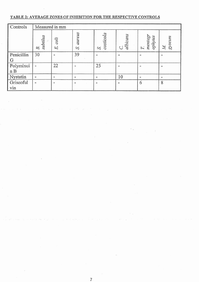
36 to 38°C for another 4 days. A standard prepared from 9mg of griseofulvin in
lmL of DMSO was also assayed for comparison purposes. Positive results were
interpreted as having an inhibition zone between the mycelium growth and the
sample disk as compared to the blank. All positive results were replicated and the
average of the diameters of the zones of inhibited microbial growth was taken.
3. Results and discussion
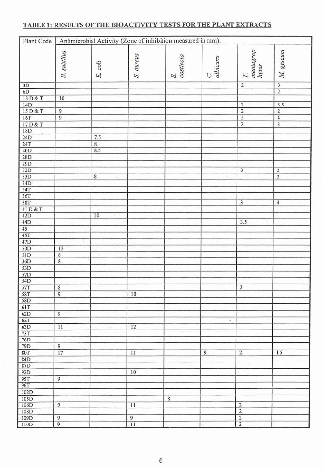
All inhibition zones of less than 1mm were recorded as negative. In each of the
assays, a standard was included to ensure that the test system was functioning
properly and to compare the activity of the sample of interests with the standard. In
addition, a positive control, which includes the solvent alone, was included, to
measure the effect, if any, of the solvent in which the samples were dissolved.
The results from the microbial assays are listed in Table I. The results of the controls
are listed in Table 2.
Approximately half of the extracts showed activity against one of the bacteria and
about a third of them against one of the fungi. Only the Endospermum macrophyllum
wood extract was active against of yeast Candida albicans. This extract was also
active against with three of the bacteria with large zones of inhibition and _both fungi.
Interestingly, the most extensive compilation of medicinal activity of Fijian plants
(Cambie and Ash, 1994) which summarises all written reports of such activity, makes
no mention of this plant. Activity is repo1ied for 450 of the roughly 2,500 plants
found in Fiji. Of course, many traditional treatments have not been documented in
writing and some of the most important ones may be family secrets.
The use of plants can also depend on availability. Many rare plants in Fiji are not
used and some not even named in the local language due to this rareness.
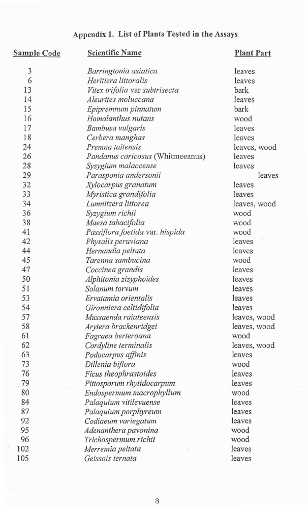
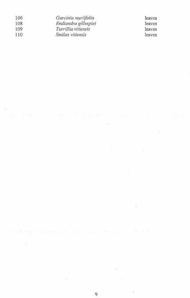
Of the forty-six plants tested eighteen are not listed in Cambie and Ash. Seventeen
are listed but the conditions for which they are reportedly used are not related to anti-
bacterial or antifungal activity. Nine plants were used for anti-bacterial purposes,
Barringtonia asiatica, Vitex trifolia var. subtrisecta, Epipremnum pinnatum, Premna
taitensis, Syzygium malaccense, Solanum torvum, Mussaenda raiateensis, Cordyline
terminalis and Dillenia bi.flora. All of these tested positive for antimicrobial activity,
and all but Barringtonia asiatica for antibacteria activity. The leaves of two plants,
Heritiera littoralis and Xylocarpus granatum, are used to treat thrush, a yeast
intection of the tongue and both displayed strong antifungal activity.
Overall only 44% of plants that did not appear in Cambie and Ash showed any
activity. For plants that were listed as used medicinally but not for antimicrobial uses
53% showed activity. As mentioned earlier, 100% of the 11 plants that were reported
4

6 Page 6

▲back to top
to be used extensively for antimicrobial purposes were also active in the laboratory.
These results indicate a good correlation between traditional use by Fijians and
activity tests in the laboratory.
One plant tested, Homalanthus nutans, has received considerable interest overseas as
it contains prostratin, a potential anti-AIDS medicine. The extract from this plant
also showed antimicrobial activity.
4. References
Bergey's manual of systematic bacteriology, Vol 1, Kreig, N. R. and Hol, J. G.
(eds), Williams and Wilkins, Baltimore, 1984.
Bergey's manual of systematic bacteriology, Vol 2, Sneath, P.H. A.; Mair, N. S.;
Sharpe, M. E. and Holt, J. G. (eds .), Williams and Wilkins, Baltimore, 1986.
Cambie, R. C. and Ash, J. (1994) Fijian medicinal plants, CSIRO, Australia,
. 365pp.
Medicinal plants in the South Pacific, WH~ Regional Publications Western
Pacific Series No. 19, 1998, 254pp.
5

7 Page 7

▲back to top
TABLE 1: RESULTS OF THE BIOACTIVITY TESTS FOR THE PLANT EXTRACTS
Plant Code Antimicrobial Activity (Zone of inhibition measured in mm).
~
:-:c-::;:
;:;
"'
Cl::i
3D
6D
13D&T
JO
14D
15 D&T
9
16T
9
17D&T
18D
24D
24T
26D
28D
29D
32D
33D
34D
34T
36T
38T
41 D&T
42D
44D
45
45T
47D
SOD
12
51D
8
36D
8
53D
57D
54D
57T
8
58T
9
58D
61T
62D
9
62T
63D
II
73T
76 D
79D
9
SOT
17
84D
87D
92D
95T
9
96T
102D
105D
106D
9
108D
109D
9
110D
9
·--a..
;~ "._:;'
;:;
'-'
<::!
kl
C,:i
7.5
8
8.5
8
10
JO
· 12
II
10
II
9
II
--~-a<.:.:!
C,:i 8"'
s"::'
<::!
u ~-'0-'
§.
!::
5°-0
-~s:: "()'.)
-E' ().)
h E:
~
~
~
2
3
2
2
3 .5
2
2
2
4
2
3
3
2
2
3
4
3.5
2
-
9
2
1.5
8
2
2
2
2
6

8 Page 8

▲back to top
TABLE 2: AVERAGE ZONES OF INHIBITION FOR THE RESPECTIVE CONTROLS
Controls Measured in mm
~
- ~ ~
~
·..~... -
..c
·~
0
(.;)
~
<:I)
:,.._
~
i::::s
c::i
~
~
~
tl'j
Penicillin 30
-
39
G
Polymixci -
22
-
nB
Nystatin -
-
-
Griseoful -
-
-
vm
~
0
·.~.\\...).
~
tl'j
0
\\.)
-
25
-
-
~
~
i::::s
u .·.~\\c.)
~
-
-
10
-
So ~
-2 ~i::::s ~
h ~ §<
-
~
~
<:I)
~~
-
-
-
-
-
6
8
7

9 Page 9

▲back to top
Appendix 1. List of Plants Tested in the Assays
Sample Code
Scientific Name
Plant Part
3
Barringtonia asiatica
leaves
6
Heritiera littoralis
leaves
13
Vitex trifolia var subtrisecta
bark
14
Aleurites moluccana
leaves
15
Epipremnum pinnatum
bark
16
Homalanthus nutans
wood
17
Bambusa vulgaris
leaves
18
Cerbera manghas
leaves
24
Premna taitensis
leaves, wood
26
Pandanus caricosus (Whitmeeanus)
leaves
28
Syzygium malaccense
leaves
29
Parasponia andersonii
leaves
32
Xylocarpus granatum
leaves
33
Myristica grandifolia
leaves
34
Lumnitzera littorea
leaves, wood
36
Syzygium richii
wood
38
Maesa tabacifolia
wood
41
Passiflorafoetida var. hispida
wood
42
Physalis peruviana
leaves
44
Hernandia peltata
leaves
45
Tarenna sambucina
wood
47
Coccinea grandis
leaves
50
Alphitonia zizyphoides
leaves
51
Solanum torvum
leaves
53
Ervatamia orientalis
leaves
54
Gironniera celtidifolia
leaves
57
Mussaenda raiateensis
leaves, wood
58
Arytera brackenridgei
leaves, wood
61
Fagraea berteroana
wood
62
Cordyline terminalis
leaves, wood
63
Podocarpus afjinis
leaves
73
Dillenia biflora
wood
76
Ficus theophrastoides
leaves
79
Pittosporum rhytidoca,pum
leaves
80
Endospermum macrophyllum
wood
84
Palaquium vitilevuense
leaves
87
Palaquium porphyreum
leaves
92
Codiaeum variegatum
leaves
95
Adenanthera pavonina
wood
96
Trichospermum richii
wood
102
Merremia peltata
leaves
105
Geissois ternata
leaves
8

10 Page 10

▲back to top
106
Garcinia myrtifolia
108
Endiandra gillespiei
109
Turrillia vitiensis
110
Smilax vitiensis
leaves
leaves
leaves
leaves
9