MSP TechRep 97-4 Digenetic trematodes of marine fishes from Suva%2C Fiji

1 Pages 1-10

▲back to top

1.1 Page 1

▲back to top
rine Sttufies
The University of the Soulh Pacific
Technical Report
DIGENETIC TREMATODES OF MARINE FISHES
FROM SUVA, FIJI. FAMILY HAPLOSPLANCHNIDAE
POCHE, 1926: DESCRIPTION OF NEW SPECIES,
A REVIEW AND AN UPDATE.
by
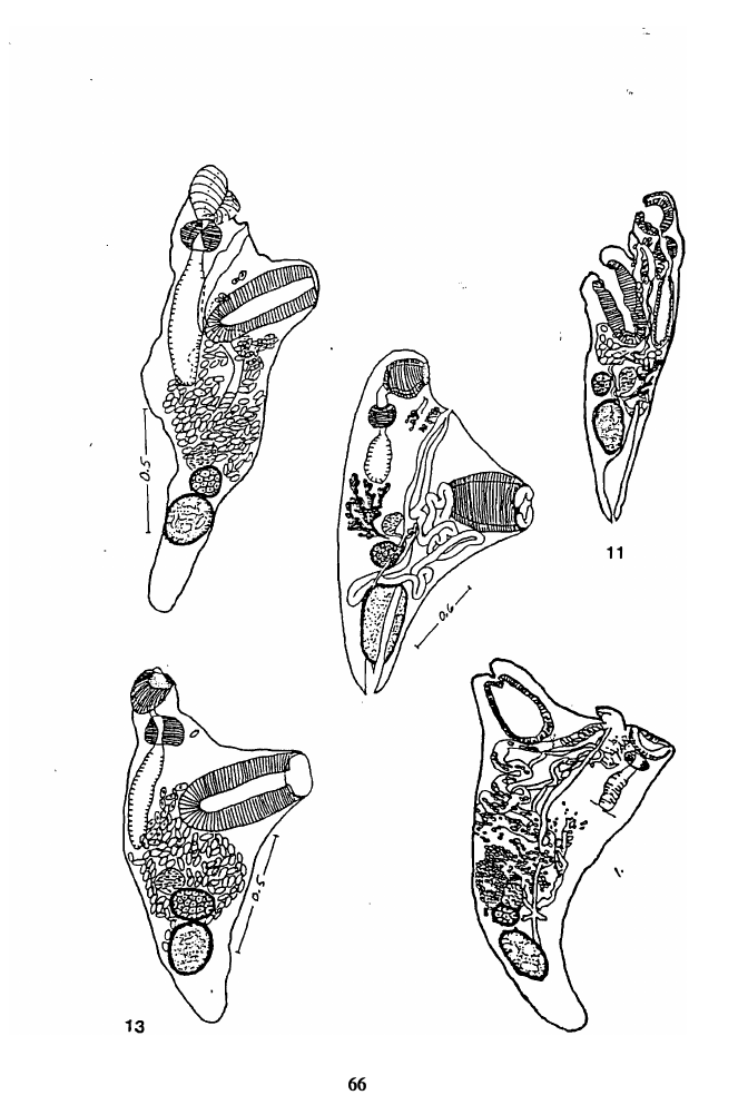
Fuad M. Nahhas
Dolores Y Rhodes
Johnson Seeto
Series
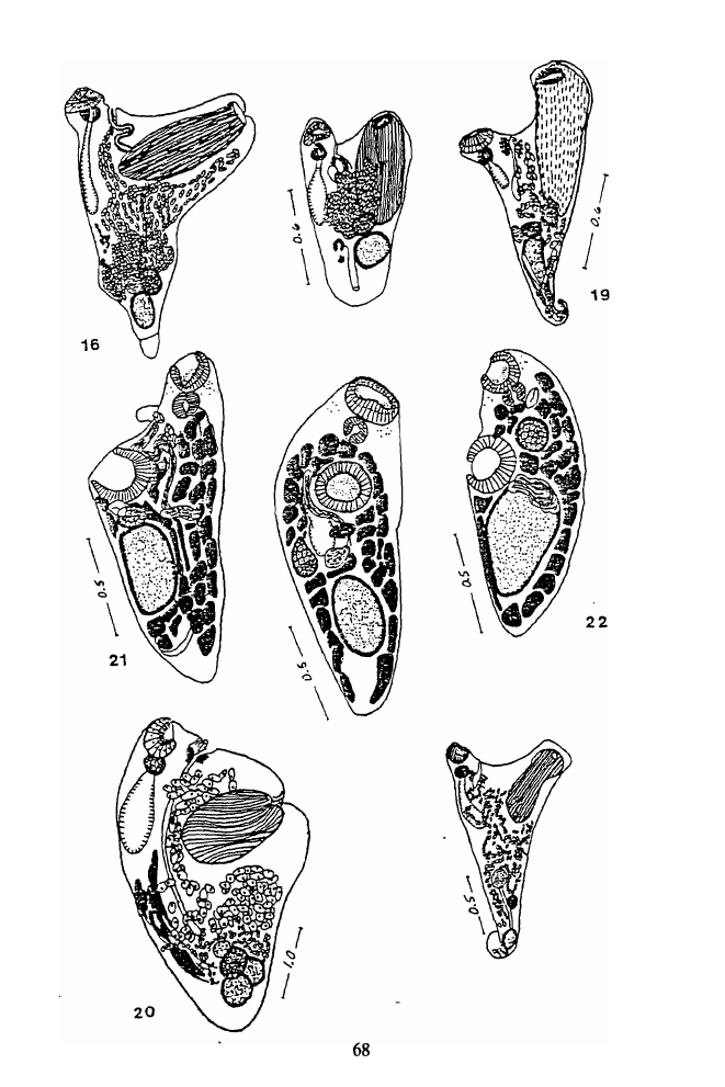
Number
ISSN 1018-2896
97/4

1.2 Page 2

▲back to top
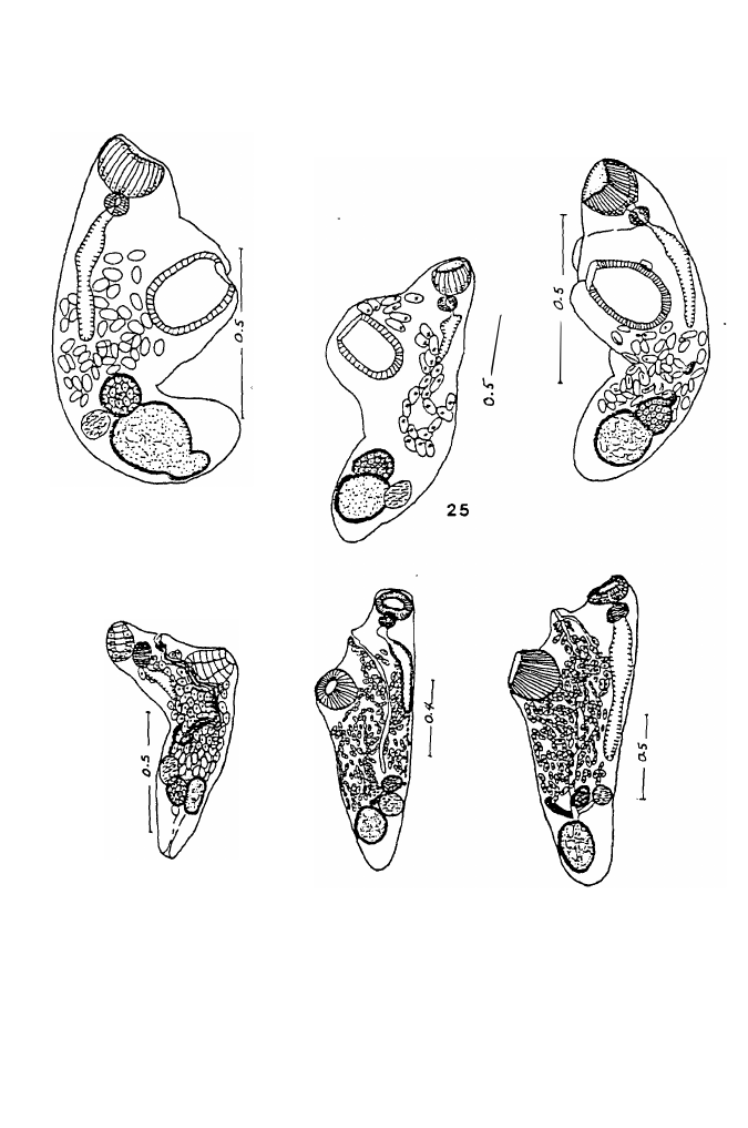
DIGENETIC TREMATODES OF MARINE FISHES FROM
SUVA, FIJI. FAMILY HAPLOSPLANCHNIDAE POCHE,
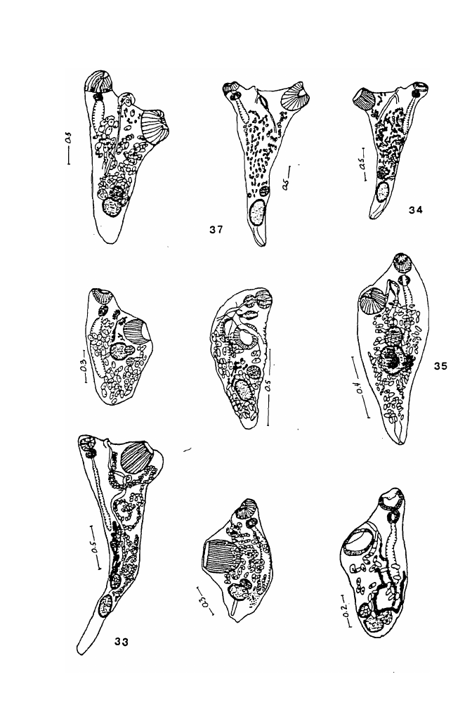
1926: DESCRIPTION OF NEW SPECIES,
A REVIEW AND AN UPDATE.
by
Fuad M. Nahhas1, Dolores Y. Rhodes1,
and Johnson Seeto2
1Department of Biological Sciences,
University of the Pacific
Stockton, CA 9521 1 , USA
2Marine Studies Programem,
The University of the South Pacific
Suva, Republic of Fiji

1.3 Page 3

▲back to top
TABLE OF CONTENTS
I. ABSTRACT .
.
.
.
.
.
.
.
.
.
.
.
.
.
.
.
.
.
.
.
.
.
.
.
.
.
.
.
.
.
.
.
.
.
.
.
.
.
.
.
.
iii
.
II. ACI<.NOWLE])CJrvfENTS . ................................ iv
III. INTRO])UCTION . . . . . . . . . . . . . . . . . . . . . . . . . . . . . . . . . . . . . . 1
IV. HISTORICAL REVIEW . . ·. . . ............................. 2
V. LIFE CYCLE ........ . ............................... 5
VI. rvtATERIALS AN]) rvtETHO])S .. . . . . , ..................... 6
VII. ])ESCRIPTION AN]) ])ISCUSSION OF THE SPECIES ............ 7
Part I - Haplosplnchnids of Fij i
1. Haplosplanchnus pachysomus ( Eysenhardt,1829) L ooss, 1902
7
2.H. purii Srivastava, 1939
16
3. Schikhobalotrema southi n. sp.
21
. 4. Schikhobalotrema sp. #1
22
5. Schikhobalotrema sp. #2
23
6. Hymenocotta mulli rvt anter, 1961
24
PART II - H aplospl anch nids of Other Parts of the W orld
7. Haplosplanchnus mugilis N ahhas and C able, 1964
25
8.H. venezuelensis Fisc hthal and N asir, 1974
26
9. H. bengalensis Ciupta and Puri, 1985
26
10. H vinodtie Ahinad, 1985
27
11. Schikhobalotrema acutum ( Linton, 1910) Skrj. and Ciusc h.,1955
28
12. S. obtusum ( Linton, 1910) Skrj.and Ciusc h., 1955
29
13. S. adacutum ( rvt anter, 1937) Skrj.and Ciusc h., 1955
30
14. S. brackyurum ( rvf anter, 1937) Skrj. and Ciusc h., 1955
31
15. S. pomacentri ( rvf anter, 1937) Skr.j and Ciusc h., 1955
31
16. S. sparisomae ( rvf anter, 1937) Skrj. and Ciusc h., 1955
32
17. S. kyphosi ( rvf anter, 1947) Skr.j and Ciusc h., 1955
33
18. S. girellae ( rvt anter and V an C leave, 1951) Skrj.and Ciusc h., 1955 34
19. S. adbrachyurum Siddiqi and Cable, 1960
34
20. S. manteri Siddiqi and C able, 1960
35
21. S. crassum Pritch ard and rvt anter, 1961
35
22. S. glomerosum Pritchard and rvt ant er, 1961
36
23. S. hawaiiensis Pritchard and rvt anter, 1961
36
24. S. robustum Pritchard and rvt anter, 1961
37
25. S. bivesiculum Nahahs and Cable, 1964
38
26. S. elongatum Nahsha and C able, 1964
39
27. S. acanthuri Yamagut i, 1970
40

1.4 Page 4

▲back to top
28. S. longivesiculatum Orecchia and Paggi, 1975
40
29. S. magnum Skinner, 1975
41
30. S. solitaria Fernandes and Goulart, 1989
42
3 1 . Pseudoschikhobalotrema heterocotylum (Nahhas and Cable, 1964)
Yamaguti, 197 1
42
32. Haplosplanchnoides hemiramphi Nahhas and Cable, 1964
43
33 . Hymenocotta manteri Overstreet, 1969
43
34 Prohaplosplanchnus diorchis Tang and Lin, 1978
44
35 . Provitellotrema crenimugilis Pan, 1984
45
36. Neohaplosplanchnus ablennis Abdul-Salam and Khalil, 1987
45
37 . Discocephalotrema kyphosi Machida, 1993
47
38. Parahaplosplanchnus cirrusaci (Lu, 1995)
48
VIII. DISCUSSION
50
IX. APPENDIX
1 . Key to the Subfamilies of Haplosplanchnidae
54
2. Key to the Genera of Haplosplanchnidae
55
3. Key to the Species of Haplosplanchnus
56
4. Key to the Species of Schikhobalotrema
57
X. TABLE 1 . Egg sire of Haplosplanchnus pachysomus-
H. caudatus complex
ro
XI. PLATES I-X
61
XII. LITERATURE CITED
81
EDITORIAL COMMITTEE FOR THIS PAPER:
R. A. Bray, Department of Zoology, The Natural .History Museum,
Cromwell Road, London, SW7 5BD, UK.
T. H. Cribb, Department of Parasitology, University of Queensland,
Brisbane, Q. 4072, Australia
ii

1.5 Page 5

▲back to top
I. ABSTRACT
Six species of haplosplanchnids are reported in this study: Haplosplanchnus
pachysomus (Eysenhardt, 1829) Looss, 1902 was found in 8 of 9 (89%) Mugil
cephalus, and H. purii Srivastava, 1939 in 1 of 8 (12. 5%) Liza vaigiensis and 1 of 9
(11%) M. cephalus; both findings represent a new locality record; Schiklwbalotrema
southi n.sp. is described from a single specimen from Scarus rivulatus. Two species
of Schiklwbalotrema are also reported but not named; Schikhobalotrema sp.# 1 from
S. rivulatus and Schikhobalotrema sp.# 2 from Acanthurus xanthopterus are described,
figured, but not named because of either damage to �e specimen or insufficient data.
Hymenocotta mulli Manter, 1961 from Mugil cephalus and Liza vaigiensis is also
reported, based on one specimen from each host (11% and 12.5% respectively).
Hop/osplanchnus caudatus (Srivastava,1939), H. straightum (Jehan,1973),H. orientalis
Gupta and Ahmad, 1979, H. stunkardi Gupta and Ahmad, 1979 and H. guptai Gupta
and. Ahmad, 1985 are considered conspecific with H. pachysomus, and H. otolithi
Gupta and Ahmad, 1979 with H. purii. Brief information on all other haplosplanchnid
species reported to date is included. Neohaplosplanchnus Lu (1995) nee Abdul-Salam
and Khalil (1987) is renamed Parahap/osplanchnus. The family Haplosplanchnidae and
its subfamilies are redefined; keys to its subfamilies, genera, and to species of
Haplosplanchnus and Schikhobalotrema are included.
Key words: Digenetic trematodes, Haplosplanchnidae, Haplosplanchnus
pachysomus, H. purii, Schiklwbalotrema southi, Schikhobalotrema sp. l,
Schikhobalotrema sp.2, Hymenocotta mulli,Parahaplosplanchnus, parasites, Mugil
cepha/.us, lil.ll vaigiensis, Sparisoma rivulatus, AcanthulUS xanthopterus,marine fishes,
Fiji.
iii

1.6 Page 6

▲back to top
II. ACKNOWLEDGMENTS
The authors are thankful to the faculty and staff of the Marine Studies
Programme (formerly Institute of Marine Resources), The University of the South
Pacific, Suva, Republic of Fiji for their assistance. Special thanks are extended to
Professor G. Robin South, Professor of Marine Studies, for making the facilities
available to the senior author to conduct this study, and Professor Rodney A. Bray of
the British Museum of Natural History, London, England, for examination of some of
the specimens, and supplying us with some literature. Professors Bray and T. H. Cribb
of the University of Queensland also served as reviewers of the manuscript; for their
valuable comments we are grateful. We thank Dr. J . Ralph Lichtenfels, Biosysternatic
Parasitology Laboratory, United States Department of Agriculture, Beltsville,
Maryland, and Professor Mary Hanson Pritchard and Dr. M. Sterner of the University
of Nebraska, Lincoln, for the loan of specimens. We also thank Christina Ninalga,
Ambre Parrish, Enjoli Gayle and Bounthavong Rasaphangthong for their assistance in
the preparation of this manuscript. The partial financial assistance of California Cedars
Products, the University of the Pacific Research Committee and the Francis R. Hunter
Fund is also gratefully acknowledged.
iv

1.7 Page 7

▲back to top
Ill. INTRODUCTION
The present paper, the third in a series (see Amin and Nahhas, 1994; Nahhas
and Wetzel, 1995) is, in part, a report of six species of haplosplanchnids, family
Haplosplanchnidae Poche, 1926, collected by the senior author between 13 January and
7 February 1992 while on sabbatical leave at the Marine Studies Programem , The
University of the South Pacific, Suva, Republic of Fiji. The Fijian species are
described in Part I of this article. Part II is a review and an update with pertinent data
on all haplosplanchnid species described to date including brief and relevant remarks.
Not included are various reports of . undescribed Haplosplanchnus and
Schikhobalotrema spp.
Two previous collections of parasites of marine fishes from the Fiji Islands have
been made, the first by Manter in 1951 (see Manter 1953, 1961, 1963a, 1963b, 1963c;
Manter and Prince, 1953), and the second, between 1979 and 1982, by the Hatsutori
Maru and other fishing boats on charter to the government of New Zealand (see Lester
et al., 1985). Manter (1961) reported one species of haplosplanchnid, Hymenocoua
mulli1 from an unidentified mullet (Mugilidae).
1

1.8 Page 8

▲back to top
IV. HISTORICAL REVIEW
The family Haplosplanchnidae, one of the smallest families of digenetic
trematodes, was established by Poche in 1926 to accommodate Haplosplanchnus
pachysomus (Eysenhardt, 1829) Loso s, 1902, a species characterized by a single cecum
and a single testis.
..
In 1910, Linton described Deradena acuta from Tylosurus marinus, D. obtusa
from Acanthurus coeruleus and Paracanthurus (Acanthurus) hepatus, and D. ovalis
from Scarus coeruleus and S. croicensis. Linton (1910) recognized a superficial
resemblance to Haplosplanchnus Looss, but noted that the vitellaria are diffuse instead
of dendritic. Manter (1931: 386) added that
"... the genus Deradena possesses a single cecum and is
therefore very similar to Haplosplanchnus Looss."
Further study by Manter (1937) led to the transfer of D. acuta and D. obtusa
to the genus Haplosplanchnus . Manter also recognized a similarity between D. ovalis
and Hapladena varia Linton, 1910 which led him to consider the former a synonym
of the latter. Manter (1937: 381) also noted
"... some constant differences between all the Tortugas
forms (H. acuta and H. obtusa) and H. pachysomus
(Eysenhardt), the type. The vitellaria are much more
extensive especially posteriorly. the prostatic cells much
less developed (unless Loso s interpreted the "neck
gland" of Linton as prostatic cells), the eggs are
definitely larger and the cecum at least somewhat
longer."
He considered these as specific rather than generic differences, redescribed H.
acutum and H. obtusum and added four new species: H. sparisomae, H. brachyurum,
H. pomacentri, and H. adacutum from various herbivorous species of fish from
Tortugas, Florida.
Srivastava (1939) described 2 new haplosplanchnids, Laruea caudatum from
Mugil (li.a)i vaigiensis from Puri, Bay of Bengal and Haplosplanchnus purii from the
same host species and locality and from Karachi, Arabian Sea. Laruea was
distinguished from Haplosplanchnus by its body shape (plump, roughly Y-shaped,
semi-spiral posterior end), the ventral sucker (long, club-shaped, tubular, more
muscular), and the position of the gonads (more anterior, just below mid-level rather
than posterior).
Manter (1947) described H. kyphosi from Kyphosus sectatrix and K. incisor
from Tortugas, Florida, and Manter and Van Cleave (19Sl) added H. girellae from
Girella nigricans from La Jolla, California.
2

1.9 Page 9

▲back to top
Skrjabin and Guschanskaja (1955) disagreed with Manter (1937) and considered
the differences, cited above, between the American species of Haplosplanchnus and H.
pachysomus to be significant enough to justify a new genus which they named
Schikhobalotrema and to which all the American species of Haplosplanchnus were
transferred. Skrjabin and Guschanskaja (1955) also did not recognire the genus Laruea
which they considered a synonym of Haplosplanchnus. They created two subfamilies,
Haplosplanchninae and Schikhobalotrematinae to accommodate Haplosplanchnus and
Schikhobalotrema respectively. Sogandares-Bemal and Sogandares ( 1 961) recognired
Schikhobalotrema as a subgenus of Haplosplanchnus.
S.
odbraScihdyduiqriuamndfrComabSlepa(r1i9s6o0m)aadsdpe. datnwdoSn. emwasnpteecriie·f�rotomthae
genus Schikhobalotrema:
"reef fish with a collar".
Pritchard and Manter (1961) added four- more: S. crassum from Stegastes fasciolatus
( = · Pomacentrus jenkenst), S. glomerosum from Acanthurus achilles and A.
sandvicensis, S. hawaiiensis from Ctenochaetus strigosus, and S. robustum from
Stegastes fasciolatus (=Pomacentrus jenkinsi), Acanthurus sandvicensis, Chaetodon
frembii and 7-ebrasoma flavescens. Manter ( 1 961) described a new genus,
Hym�npcotta, to accommodate H. multi from an unidentified mullet (family
Mugilidae). Nahash and Cable (1964) described Schkikhobalotrema bivesiculum from
Abudefduf saxatilis, S. elongatum from Mugil cephalus and M. curema, and S.
heterocotylum from Pseudoscarus guacamaia. They also reported the first species of
Haplosplanchnus from the Caribbean which they named H. mugilis from M. curema,
and described an amphistomatous haplosplanchnid from Hemiramphus brasiliensis
which they called Haplosplanchnoides hemiramphi. Overstreet ( 1969) added a second
species to the genus Hymenocotta, H. manteri, from M. cephalus from Biscayne Bay,
Florida. Yamaguti ( 1 970) described Schikhobalotrema acanthuri from Acanthurus
sandvicensis, A. nigrofuscus, and 7-ebrasoma flavescens.
Yamaguti ( 1 97 1 ) revised the family recognizing the genus Haplosplanchnus
with 3 species and Schikhobalotrema with 17. He rejected Skrjabin and Guschanskaja's
synonymy of Laruea with Haplosplanchnus. He also transferred Schikhobalotrema
heterocotylum Nahhas and Cable, 1964 to a new genus Pseudoschikhobalotrema, and
Hymenocotta manteri Overstreet, 1 969 to Hymenocottoides.
Zhukov (1971 ) described Haplosplanchnus bivitellosus from Mugil cephalus and
M. soiuy from the basins of the Japan and Yellow Seas. In 1973, Jehan described
Laruea straightum from Pseudosciaena diacanthus and Rachycentron canadus.
Fischthal and Nasir ( 1974) added H. venezuelensis from Antennarius multiocellatus
from Los Roques Islands, Venezuela. Orecchia and Paggi ( 1 975) described
Schikhobalotrema longivesiculatum from Blennius sanguinolentus from the Gulf of
Gaeta, Italy. Skinner ( 1 975) added S. magnum from M. cephalus from Biscayne Bay,
Florida. Tang and Lin ( 1978) described Haplosplanchnus cuneatus and H. elongatus
from M. cephalus, and Prohaplosplanchnus diorchis from Liza haematochilus and M.
cephalus all from the Min River, Fujian, China. Gupta and Ahmad (1979) described
Haplosplanchnus indicus from M. vaigiensis, H. orientalis from llisha elongata
( =Pellona elongata), H. otolithi from Otolithus ruber, and H. stunkardi from
3

1.10 Page 10

▲back to top
Acanthurus sandvicensis all from the Bay of Bengal, at Puri, Orissa. Pan (1984) added
a new genus and species Provitellotrema crenimugilis from Crenimugil crenilabis.
Ahmad (1985) described Haplosplanchnus guptai and H. vinodae from M. cephalus
from the Panjim Coast, Goa, India. Gupta and Puri (1985) described H. bengalensis
from Acanthurus sandvicensis from the Bay of Bengal, at Puri , Orissa. Abdul-Salam
and Khalil (1987) added a new genus and species, Neohaplosplanchnus ablennis, from
Ablennes hians from the Kuwaiti coast, Arabian (Persian) Gulf. Fernandes and
Goulart(1989) described Schikhobalotrema solitaria from Stephanolepis hispidus from
Praia de Copacabana, Rio de Janeiro; Machida (1993) added a new genus
Discocephalotrema to accommodate D. kyphosi from Kyphosus cinerascens and created
for it a new subfamily Discocephalotrematinae; Lu (1995), unaware of the description
of Nephaplo-splanchnus ablennis Abdul-Salam and Khalil, 1987 from Kuwait, used the
same generic name as a new genus to accommodate N. cirrusaci from Engraulis
japonica from Shallow Sea, Guandong Province, China.
4

2 Pages 11-20

▲back to top

2.1 Page 11

▲back to top
V. LIFE CYCLE
Two life cycles have been described in the family Haplosplanchnidae Poche,
1926. Cable (1954) described a partial life cycle involving sporocyst stages within the
marine snail Cerithium variable (Gastropoda, Cerithiidae), the development of the
cercariae within the snail and their encystment on vegetation in water as metacercariae.
Whether the haplosplanchnid egg is eaten by the snail or hatches in water and later
penetrates the snail intermediate host was not determined. A redial stage is apparently
absent. Cable (1954) suggested that these developmental stages were most likely the
larval forms of Schikhobalotrema acutum (Linfon, 1910) Skrjabin and Guschanskaja,
1955, the most common haplosplanchnid in Caribbean and adjacent waters.
Fares and Maillard (1975) d.escribed the life cycle of Haplosplanchnus
pachysomus (Eysenhardt, 1829) Looss, 1902. They had found natural infections in Liza
auratus, L. ramada, Mugil cephalus and Chelon labrosus, and haplosplanchnid
cercariae emerging from naturally infected Hydrobia ventrosa (Gastropoda:
Hydrobiidae) in littoral waters of Languedoc-Roussillon, French Mediterranean coast.
Fares and Maillard used laboratory-raised H. ventrosa snails, and mullets that had bene
collected at a young age and kept in aquaria, to demonstrate a complete life cycle.
Their experiments showed that ciliated miracidia, which had emerged from the uterus
of adult H. pachysomus, penetrate the snails, develop in the gonads into sporocysts
which give rise to rediae which, in tum, produce cercariae. The phototropic cercariae
swim to the surface and encyst as metacercariae in the surrounding environment (milieu
ambient). Only 4-5 day old metacercariae were infective, which when eaten by mullets,
develop into adults in the anterior part of the intestine of the fish. Fares and Maillard
(1975) also gave a detailed description of the anatomy of the adult.
Recently Abdul-Salam and Sreelatha (1995) described Cercaria kuwaitae VI,
y a new haplosplanchnid cercaria which develops in sporocysts in Cerithidea cingulata.
The onl known adult haplosplanchnid from the Arabian Gulf is Neohaplosplanchnus
ablennz's Abdul-Salam and Khalil, 1987.
5

2.2 Page 12

▲back to top
VI. MATERIALS AND METHODS
A total of 236 fishes were obtained from several sources in Laucala Bay, Suva
including traps, nets, spear fishing, and commercial fishermen. Except for a few fishes
that were purchased, all were captured live on reefs and lagoons of Laucala Bay, Suva,
a few miles from the laboratory of the Marine Studies Programme (formerly Institute
of Marine Resources). Fifty species, representing 32 genera and 20 families were
collected. Four species: Acanthurus xan thop terus (Acanthuridae), Li za vaigiensis and
Mugil cephalus (Mugilidae), and Scarus rivulatus (Scaridae), harbored
haplosplanchnids. The fish were kept alive in tanks until shortly before examination.
After removal from the host, the digeneans were washed in 0.7% saline, many studied
alive before they were fixed with alcohol-formalin-acetic acid (A.F.A.) under slight
coverslip pressure. The worms were then transferred to a dish, left in the fixative
overnight, then stored in 70% ethanol. After washing with three changes of 50%
isopropanol, the specimens were stained with Semichon's acetocarmine (Carolina
Biological Supply Company), destained in 0.5% acid alcohol, dehydrated in ascending
series of isopropanol, cleared in methyl salicylate, rinsed in xylol, and mounted in
Kleermount (Carolina Biological Supply Company).
Measurements are expressed in millimeters except for eggs which are in
micrometers (µm). Sucker ratio was calculated from the mean of the length (depth) and
width and is expressed with the oral sucker taken as 1. Drawings of specimens obtained
in this study were prepared by microprojection and details filled in through microscopic
observations. Drawings of other species were made by tracing original diagrams. One
asterisk* indicates a new synonym. The number of specimens recovered from each
infected fish and the number of fish examined are indicated next to each host species
listed in the description of the Fijian species. Discussions or remarks, where necessary,
are also presented with each species.
Holotypes are deposited in the Parasite Collection of the United States National
Parasite Collection (USNPC), Beltsville, Maryland; voucher specimens of some species
are in the British Museum of Natural History, BM(NH), London, and Harold W .
Manter Laboratory (HWML), University of N ebraska State Museum, Lincoln. Fishes
were identified by the third author, Johnson Seeto. References used included an
unpublished manuscript on fishes of the Fiji Islands, Nelson ( 1984), Myers ( 1989), and
Randall et al. (1990).
6

2.3 Page 13

▲back to top
VII. DESCRIPTION AND DISCUSSION OF THE FIJIAN SPECIES
Part I - Haplosplnchnids of Fiji
GENUS HAPLOSPLANCHNUS Looss, 1902
To date 16 species of Haplosplanchnus have been reponed; four of these are
listed in Yamaguti ( 197 1): H. pachysomus (Eysenhardt, 1829) Looss, 1902, H.
caudatus (Srivastava, 1939) listed as Laruea caudatum, H. purii Srivastava, 1939 , and
H. mugilis Nahhas and Cable, 1964 . Twelve others were described after 197 1 , and are
b19ri7e3t1)yAdhismcuasds,ed19i8n5t,hHis .pvaepneer:zuHel.ebnisvisiteFliloscshutsh· aZlhaunkdo.vN, a1s9ir7,
1, H.
1974,
straightum (Jehan,
H. cuneatus Tang
and Lin, 1978, H. elongatus Tang and Lin, 1978, H. indicus Gupta and Ahmad, 1979 ,
H. orientalis Gupta and Ahtnad, 1979, H. otolithi Gupta and Ahtnad, 1979, H.
stunkardi Gupta and Ahmad, 1979 , ii. bengalensis Gupta and Puri, 1985, H. guptai
Ahmad, 1985, and H. vinodae Ahmad, 1985 .
There seems to be a correlation between the presence or absence of muscular
acetabtilar lips, the ratio of the oral sucker to the ventral sucker, and the length-width
ratio of the ventral sucker. The 16 species of HaplosjJlanchnus can be separated into
two groups. Group 1 is characteriz.ed by the presence of muscular acetabular lips, a
. ventral sucker at least twice the oral sucker and its length at least twice its width. Six
species are included in this group: H. pachysomus, H. caudatus, H. straigh tum, H.
orientalis , H. stunkardi, and H. guptai. Group 2 is characteriz.ed by absence of
muscular acetabular lips, a sucker ratio of 1 :2 or less, and ventral sucker length not
much greater than its width. This group consists of 10 species: H. purii, H. mugilis,
H. bivitellosus, H. venezuelensis, H. cuneatus, H. elonga tus, H. indicus, H. otolithi,
H. bengalensis, and H. vinodae.
Two of the 16 species, H. pachysomus and H. purii were encountered in this
study. ' ,
1.
Haplosplanchnus pachysomus (Eysenhardt, 1829) Looss, 1902
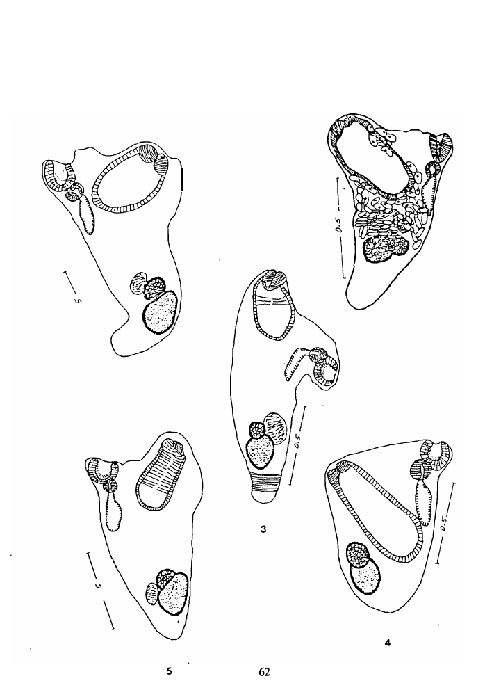
Figs. 1-5 Plate I
Figs. 1 1- 15 Plate III
SYNONYMS:
Distoma pachysoma Eysenhardt, 1829
Podocotyle pachysomus (Eysenhardt, 1829)
Stossich, 1898
* Laruea caudata Srivastava, 1939
*L. straightum Jehan, 1973
*Haplosplanchnus caudatus Srivastava, 1939
*H. straightum (Jehan, 1973) Ahmad, 1985
*H. orientalis Gupta and Ahmad, 1979
*H. stunkardi Gupta and Ahmad, 1979
*H . guptai Ahmad, 1985
7

2.4 Page 14

▲back to top
HOST:
Mugil cephalus (Mugilidae)
3/4/8/8/9/11115/19 in 8 of 9
SITE:
Intestine
DATE O F
COLLECTION : Jan.19, 1992
Feb. 3, 1992
DEPOSITE D
SPECIMENS : USNPC 83982
BM(NH)l993.4.2.81 . 84
HWML 37842
Description (based on 77 specimens, including several live ones; measurements
on 16). Body brownish-yellow in life, Y-shaped or somewhat triangular, tapering
posteriorly, ventrally rolled up in some, dorsally in others, and rounded in a third
group, 0.800-1. 200 long, 0.475-0.875 wide at acetabular level. Cuticle aspinose, eye­
spot pigment diffuse. Oral sucker ovoid, 0. 110-0. 200 long, 0.089-0.240 wide; ventral
sucker elongated, 0.450-0 . ()()() long, 0. 180-0.370 wide, with two muscular lips,
situated in the longer arm of the Y-shaped body; ventral sucker length (depth) 2-4
times width. Prepharynx shon; pharynx ovoid, 0. 073-0.120 long, 0. 070-0. 110 wide;
esophagus shon; cecum 0. 140-0.400 long, extending to posterior level of ventral
sucker. Testis single, ovoid, 0 . 140-0. 310 long, 0. 100-0.250 wide in posterior half of
body; seminal vesicle tubular, extending to anterior level of ventral sucker; prostatic
duct surrounded by prostate cells; ejaculatory duct shon. Ovary globular or ovoid,
anterior or anterolateral to, often contiguous with testis in fixed specimens, 0. 070-
0.190 long, 0. 070-0. 140 wide; seminal receptacle variable in position and size, usually
anterolateral to, smaller, same size, or larger than ovary, 0. 078-0.250 long, 0.050-
0.180 wide; vitelline follicles 10-15, poorly developed, distributed from ovary to near
posterior level of ventral sucker. Uterus variable in its posterior extent, usually to
anterior level of testis, and in a few, extending beyond its posterior level; eggs
containing developing miracidia with prominent eye-spots, 45-67 long, 27-35 wide;
Genital atrium tubular; genital pore midway between oral sucker and ventral sucker.
Excretory bladder Y-shaped or sac-like with wide arms, containing a large concretion­
like structure, with several smaller ones in one or both of the excretory arms.
H. pachysomus has been reported by several authors (see Bray, 1984) and from
various species of mullets belonging to the Mugil-Liza group, Crenimugil crenilabis,
artd Chelan labrosus (Mugilidae).
Several characteristics have bene used to distinguish the six species of
Haplosplanchnus with an elongated ventral sucker (group 1). These included body
shape, topography of the gonads, whether the gonads are contiguous or separated from
each other, extent of the uterus relative to the testis, size and position of the seminal
receptacle in relation to the ovary, length of the cecum, number and distribution of the
vitellaria, nature of the prostatic complex, and egg size.
8

2.5 Page 15

▲back to top
The Fijian specimens were initially identified as H. caudatus. It was noted,
however, that they also have many similarities to H. pachysomus; furthermore, when
the various reports of H. caudatus and H. pachysomus were reviewed, it became
evident that there is complete overlap between the two species not only in terms of
organ topography but also in measurements. It was also noted that there are very few
reports in which the two species were compared. The following historical review and
comparison led to the conclusion that H. caudatus is not distinct from H. pachysomus;
furthennore, several other species (H. straightum, H. orientalis, H. stunkardi, and H.
guptm) should also be considered synonyms of H. caudatus and by extension of H.
pachysomus.
·
A brief review of these species follows:
Haplosplanchnus caudatus (Srivastava, 1939) Skrj. and Gusch., 1955
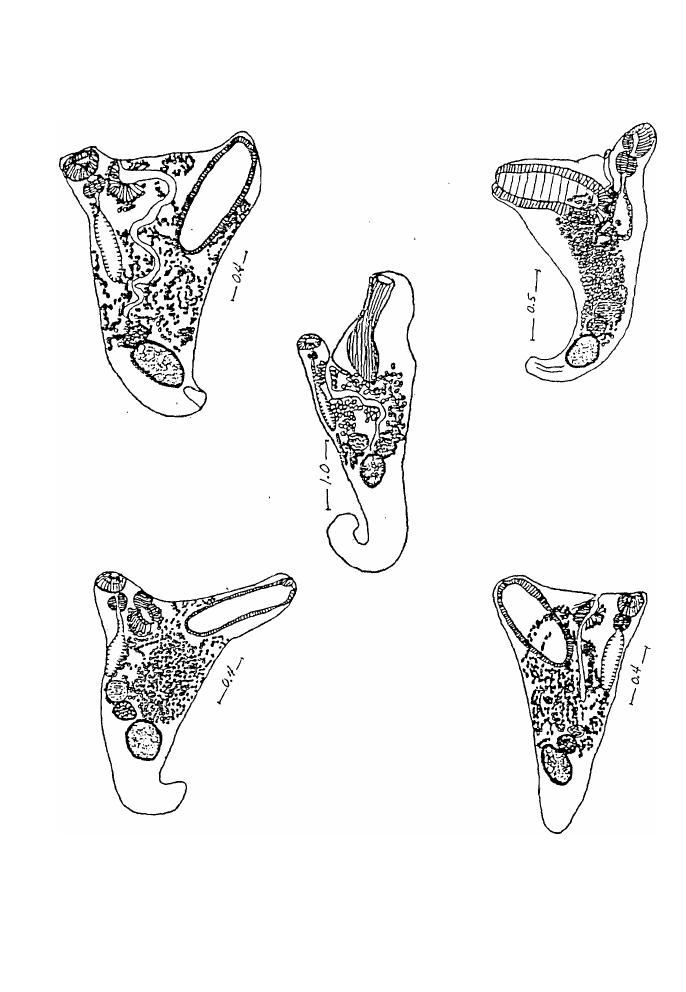
Figs. 6-10 Plate II
SYNONYM: Laro.ea caudatum Srivastava, 1939
Three reports describe or discuss H. caudatus. Srivastava collected 3 specimens
from Mugil (Liza) vaigiensis Quoy arxl Gaimard, at Puri, Bay of Bengal in 1935, gave
a brief description in 1937 and a more detailed one in 1939 under the generic name
Laruea. In comparing the new genus to Haplosplanchnus, Srivastava (1939: 70) stated
"The new genus, Laruea differs remarkably from
the type genus in the peculiar shape of its body
and of the ventral sucker, and in the position of
its gonads and vitellaria. In all other important
features the two genera are alike."
Madhavi (1979: 239) stated that her 14 specimens from the same host species
and from M. cephalus from Waltair coast, Bay of Bengal
"... showed considerable variation in body shape. The
fully gravid specimens have a shape identical to that
represented for type specimen. They have a
characteristic Y-shaped body with a curved tail like
structure near posterior end arxl the long disc like ventral
sucker situated on a lateral extension. In some specimens
the posterior tail like portion retracted into the body
proper and such specimens had a blunt posterior end.
The younger specimens had a diff�rent body shape which
is almost triangular without any tail like portion near
posterior end. The specimens also showed variation in
the extent of the cecum, some had a longer cecum
almost reaching the level of ovary, in others it is much
9

2.6 Page 16

▲back to top
smaller. Eggs measured 39 to 43 in length and 18 to 23
in width."
Madhavi accepted the synonymy of Laruea with Haplosplanchnus as suggested
by Skrjabin and Guschanskaja (1955) and concluded that
" ... the present observation also supports this view as it
is clearly seen that the body shape is variable, and cannot
be used as a reliable taxonomic character. The only
feature that separated the two genera is the shape of the
ventral sucker which has no more than any specific
significance. "
The 77 Fijian specimens obtained from eight M. cephalus, ranging in sire from
10-15 centimeters and captured on two different dates, are in agreement with the
several descriptions and measurements of both H. pachysomus and H. caudatus as
reported in the literature. The Fijian material shows similar variations as described by
Madhavi for H. caudatus. The elongated, curved posterior end described by Srivastava
(1939) was seen in the live material when the worms were placed in a dish on ice and
became fully stretched. When the worms were fixed at room temperature, that shape
changed to triangular, roughly Y-shape, or elongate as seen in figures 1-5 (Plate I).
Many worms took a circular fonn when the posterior end was grabbed by the powerful
muscular acetabular lips of the ventral sucker.
Figures 1, 2, 3, and 5 also show variations in the si.ze and location of the
seminal receptacle. The gonads and the seminal receptacles are usually close together,
but their position in the body depends on degree of contraction of the worms and the
shape they take.
Neither Srivastava (1939) nor Madhavi (1979) described muscular acetabular
lips on the ventral sucker of H. caudatus. Ahmad (1985) had requested Dr. Madhavi
to reexamine her slides of H. caudatus for the presence of these structures, and she
confirmed their presence. Figures 1-5 show the muscular acetabular lips and the
different shapes that the ventral sucker may take as a result of its contraction,
extension, and to a lesser extent influence of cover slip pressure during fixation.
H. caudatus was also reported by Bray (1984) from M. cephalus from
Sodwana, Natal, South Africa. He gave no detailed description or measurements, but
at our request (personal communication: FMN) he measured and reported egg siz.e of
42-48 X 26-28 µm.
10

2.7 Page 17

▲back to top
Bray (1984: 278) noted that:
" . . . it (H. caudatus) is very similar to the type of the
genus, Haplosplanchnus pachysomus (Eysenhardt, 1829)
Looss, 1902 and may be synonymous. . . the difference
between H. caudatus and H. pachysomus is in the degree
of development of the ventral sucker for although
elongate in both species, this condition is apparently
greatly more exaggerated in H. caudatus. "
His figures of H. caudatus as well as those of Srivastava and Madhavi are shown in
figures 6-10 (Plate II). Bray (1984) compared the ventral sucker of H. caudatus with
H. pachysomus as figured by various authors including that of Stossich which shows
" .. . an elongate sucker similar to that of H. caudatus,
although the specimen is from the Mediterranean."
This suggested to him
, " .. :that both species occur in the Mediterranean or that
there is but one variable species. "
Dr. Bray examined six specimens we sent him and he was in agreement with
our preliminary identification of the Fijian specimens as H. caudatus. However, further
review of the literature led us to reconsider our original identification and to conclude
that the two species are conspecific. Bray (1984) al8o pointed out the similarity of
Laruea straightum Jehan, 1973, H. orientalis Gupta and Ahmad, 1979, H. stunkardi
Gupta and Ahmad, 1979, all from non-mullets, to H. caudatus, and suggested that they
may be synonyms of the latter. To this group, which we shall call the 'H. caudatus
complex, we add H. guptai Ahmad, 1985 from M. cephalus. All these species,
including ,L. straightum, which was later transferred to Haplosplanchnus by Ahmad
(1985),. are known to have muscular acetabular lips, and their body size, internal
organs, and other measurements overlap. Their eggs are fully developed with eye­
spotted miracidia. The variations that we have seen, during live observations and in
fixed specimens, lead us to agree with Bray's insight and suggestion. A brief sumam ry
of each and the justification given by their authors follow.
Haplosplanchnus straightum (Jehan, 1973) Ahinad, 1985
Fig. 16, Plate IV
SYNONYM: Laruea straightum Jehan, 1973
Specimens of this species were collected from Pseudosciaena diacanthus
(Sciaenidae) and Rachycentron canadus ( Rachycentridae).
11

2.8 Page 18

▲back to top
The similarity of H. straightum to some Fijian specimens ( Figures 3 and 5,
Plate I) is quite evident. H. straightum is considered a synonym of H. caudatus, and
by extension, of H. pachysomus.
Haplosplanchnus orientalis Gupta and Ahmad, 1979
Fig. 17 Plate IV
H. orientalis, described from a single specimen taken from one /lisha elongata
( =Pellona elongata) (Clupeidae) out of 53 examined from Puri, Orissa, Bay of Bengal,
is probably the same species reported by Gupta and Ahmad in 1975 as Laruea
orientalis.
Gupta and Ahmad ( 1979:187) stated that
"the new form has a close resemblance with H.
caudatum Srivastava, 1939 in having ventral sucker
long, club-shaped and genital pore at bifurcation of Y
but differs from it in having testis, ovary and
receptaculum seminis separated from each other by
uterine coils instead of close together, in having testis
near hind end of body instead of behind first half of
body, in the extension of uterus up to hind end of body
instead of up to testis, in the extension of intestinal
cecum up to a little posterior to ventral sucker instead of
up to a little anterior to receptaculum seminis, in the
extension of vitellaria between ovary and blind end of
intestinal cecum instead of between testis, the ovary and
blind end of intestinal cecum and in having ovary in
posterior third of body instead of in middle third of
body. Accordingly it is regarded as a new species with
the specific name H. orientalis sp. n. "
Since this description is based on a single specimen, the constancy of these
characteristics is hard to determine. The Fijian specimens exhibit variations in all the
characteristics cited by the authors. Bray ( 1984) suggested that differences involving
the distribution of the uterus
" may be of little importance and, in geographical terms,
an intermediate form may occur. "
In view of M adhavi's findings and the variations seen in the Fijian material, H.
orientalis is reduced to a synonym of H. caudatus and by extension of H. pachysomus.
12

2.9 Page 19

▲back to top
Haplosplanchnus stunkardi Gupta and Ahmad , 1979.
Fig. 18 Plate IV
H. stunk:ardi was described from two specimens in one Acanthurus sandvicensis
out of 4 1 examined at Puri.
Gupta and Ahmad ( 1979: 188)) acknowledged its similarity to H. caudatus
stating that
"the new form has a close resemblance with H.
caudatum Srivastava, 1939 and H. orjentalis sp. n. in
having long, club-shaped ventral sucker and genital pore
at bifurcation of Y but differs from both these forms in
the extension of ventral.sucker posteriorly up to anterior
to testis, in the extension of intestinal cecum up to
middle of ovary, in the extension of vitellaria between
middle of testis and blind end of intestinal cecum and in
having posterior end rounded. The new form further
. differs from H. caudatum in the extension of uterus
between middle of testis and middle of ventral sucker
instead of between testi s and ventral sucker. The new
form can further be distingui shed from H. orientalis in
having testis and ovary close together instead of
separated from each other by uterine coils and in the
extension of uterus between middle of testis and middle
of ventral sucker instead of between hind end of body
and one third length of ventral sucker. Accordingly it is
regarded as a new species with the specific name H.
stunkardi sp. n."
The characteristics cited by Gupta and Ahmad as distinguishing features of H.
stunkardi are evident in several of the 77 Fijian specimens particularly in the length of
the ventral sucker relative to total body length . Gupta and Ahmad's figure of H.
stunkardi reproduced in figure 18 (Plate IV) is clearly very similar to a Fijian specimen
shown in figure 4 (Plate I). We consider this species also a synonym of H. caudatus,
and by extension, a synonym of H. pachysomus.
H. stunkardi was also reported, but not described or figured, by Ezz El-Dien
et al. ( 1990) from M. cephalus and M. chelo from the Suez Canal , Egypt. These
authors also indicate that Al-Bassel found it in M. capito at Lake Quarun, Nile River
in 1987.
13

2.10 Page 20

▲back to top
Haplosplanchnus guptai Ahmad, 1985
Fig. 19 Plate IV
This species was described from nine specimens from the intestine of M.
cephalus from the Panjim Coast, Goa, India.
According to Ahmad (1985: 193)
"the new species most closely resembles H. caudatum
(Srivastava, 1939) , H. straightum (Jehan, 1973) , H.
orientalis Gupta and Ahmad, 1979 and H. stunkardi
Gupta and Alunad, 1CJ79 in the shape of body and in the
structure of ventral sucker. The new species differs from
H. caudatum, H. straightum and H. stunkardi in the
extension of uterus posteriorly up to hind end of body
instead of uterus extending only up to anterior margin of
testis. The new species closely resembles H. orientalis in
the extension of uterus up to posterior end of body but
differs from it in having gonads close together instead of
well separated from one another, in having seminal
receptacle lateral to ovary instead of anterior to ovary, in
having testis elongate and of larger size instead of testis
oval and of smaller size, in having seminal receptacle
contiguous with ovary instead of well anterior to it, in
having eggs of larger size instead of smaller size (35-42
X 18-20 µm) , vitellaria consisting of 7-10 follicles,
postovarian, grouped together instead of vitellaria
consisting of numerous small follicles which are
confined between ovary and blind end of cecum, in
having seminal vesicle of smaller size and stopping much
anterior to testis instead of seminal vesicle of larger size
and stopping near testis."
The only feature that was not observed in the Fijian material is an elongated
testis. This characteristic was emphasized by Ahmad as separating H. guptai from H.
orientalis. In some Fijian specimens, the uterus extends slightly posterior to the testis
but not to the posterior end of the body. The larger eggs (50-65 X 26-37) of H. Guptai
are within the range found in the Fijian specimens, and those given for H. pachysomus.
They also represent an intermediate range between the H caudaJus complex and H.
pachysomus (Table I). We do not consider the shape of the testis significant enough to
justify a new species; in all other respects it is similar to H. pachysomus.
In contrast with the few records on H. caudaJUS, there have bene at least 12
reports of H. pachysomus in recent years from mullets of the Adriatic , Black, and
Mediterranean seas, and the Nile River.
14

3 Pages 21-30

▲back to top

3.1 Page 21

▲back to top
Yamaguti (1934:361)) was the first to report H. pachysomus from outside these
bodies of water. He recovered this species from young and adult M. cephalus from the
Inland Sea of Japan, stating:
"This species has been dealt with by Looss with
such accuracy that there is no need of redescription, but
I may take this opportunity to supply some details which
are missing in Looss' description.
In the first place the living worm is brownish
yellow throughout, although the alcoholic specimens are
entirely colorless.
The excretory vesicle is divided into two parts;
the posterior is an elongated sac extending to near the
testis, but the anterior is relatively narrow and bifurcates
just behind the testis into lateral arms passing forwards
along the lateral borders of the testis.
The vas deferens forms an elongate oval vesicula
seminalis at its distal end. The terminal portion of the
uterus is distended with embryonated eggs, some hatched
in . the uterus. The empty shells in the uterus measure
about
0.051
X
0.027
mm
.
The
elliptical
free
miracidia
are 0.06-0.07 X 0. 027 -0. 03 mm and covered with short
cilia all over; they have a large x-shaped eye-spot with
a lens on each concave side; at the extreme anterior end
there is a very small sharply pointed stylet"
It should be ooted that neither Srivastava (1939) nor Madhavi (1979) made any
reference to Yamaguti's 1934 report of H. pachysomus from Japan. The presence of
muscular lips in H. pachysomus (Figure 11, Plate Ill) was not described by Looss but
several investigators including Fares and Maillard (1975) reported them (Figure 14,
Plate Ill). Our examination of Fischthal and Kuntz's specimens (Figures 12 and 13,
Plate III) deposited at USNPC also confirms their presence.
Loso s's description of this species was based on specimens from M. cepha.lus
and M. chelo from Trieste, the Adriatic Sea. In addition to the reports of Fischthal and
Kuntz (1963) from M. cephalus and M. (Liza) ramada from Egypt, and Fares and
Maillard (1975) from Liza aurota, L. ramada, M. cephalus and Che/on labrosus from
the French Mediterranean coast, there were several others including Papema ( 1964)
from M. cephalus, M. auratus, M. labeo, and M. chelo from Israel, from the Nile
River and the Mediterranean by several Egyptian parasitologists and in the last 1O years
those of Garippa et al. (1988) from M. cephalus in Italy, and Brglez and Paradiznik
(1988) from M. cephalus in the Adriatic. H. pachysomus was also found in L. ramoda
by Orecchia et al. (1988) in the Mediterranean, and by Radujkovic et a/.(1989) and
Radujkovic and Raibaut (1989) in the Adriatic. Gaevskaya and Dmitrieva ( 1992)
redescribed H. pachysomus from M. cephalus, L. ourata and L. saliens from the Black
Sea; their report and that of Bray ( 1984), as far as we can determine, are the only two
15

3.2 Page 22

▲back to top
that compared H. pachysomus with H. caudatus. Bray's conclusion, that the only
difference between the two species is the degree of development of the ventral sucker,
has already been referred to. Gaevskaya and Dmitrieva, on the other hand,
distinguished the two species by egg size which is somewhat greater for H. pachysomus
( 39-57 x 1 8-30) than for H. caudotus ( 39-43 x 18-22). Table I lists egg size range for
H. pachysomus-H. caudatus complex as reported in the literature. The table suggests
that eggs from the Adriatic, Black, and Mediterranean specimens of H. pachysomus
tend to have a slightly larger size compared with those of the H. caudatus complex of
the Indian, southern Atlantic oceans and the Arabian Sea, but the overlap is clear. On
the basis of egg size, the Fijian material must be identified as H. pachysomus; when
shape, topography of organs, and suckers are considered, the only conclusion that can
be made, to paraphrase Bray ( 1984), is either all six species occur in Fijian waters or
there is but one variable species.
In group 2 of the genus Haplosplanchnus no muscular acetabular lips are
present, the ventral: oral sucker ratio and the ventral sucker length compared to its
width is less than 2: 1 . This group includes H. purii Srivastava, 1939, H. mugilis
Nahsha and Cable, 1964, H. bivitellosus Zhukov, 197 1 , H. venezuelensis Fischthal and
Nasir, 1974, H. cuneatus Tang and Lin, 1 978, H. elongatus Tang and Lin, 1978, H.
indicus Gupta and Ahtnad, 1 979, H. otolithi Gupta and Ahmad, 1979, H. bengalensis
Gupta and Puri, 1 985, and H. vinodae Ahmad, 1985.
2. Haplosplanchnus purii Srivastava, 1939
Figs. 24-29 Plate V
SYNONYMS:
H. bivitellosus (Zhukov, 1 97 1) Bray, 1984
H. cuneatus (Tang and Lin, 1978) Bray, 1984
H. elongatus (Tang and Lin, 1 978) Bray, 1984
H. indicus (Gupta and Ahmad, 1 979) Bray, 1 984
*H. otolithi Gupta and Ahmad, 1 979
HOSTS:
Liza vaigiensis (Mugilidae) 7/l of 8
Mugil cephalus (Mugilidae) 11 1 of 9
SITE:
Intestine
DATE OF
COLLECTION: Jan. 19, 1992
Jan. 3 1, 1992
DEPOSITED
SPECIMENS: USNPC 83983
BM(NH) 1994.6. 14. 1
HWML 37843
Description and measurements based on eight specimens. Body somewhat Y­
shaped, rounded posteriorly, 0.725- 1.425 long, 0.400-0.650 wide at acetabular level.
Cuticle aspinose, eye-spot pigment diffuse. Oral sucker ovoid, 0. 1 10-0. 1 90 long,
16

3.3 Page 23

▲back to top
0.150-0.200 wide; ventral sucker without acetabular lips, somewhat cylindrical, 0.250-
0.350 long, 0.170-0.230 wide; sucker ratio 1:1.6-1.8 ; ventral sucker length ( depth):
width about 1.5:1. Prepharynx short; pharynx spherical, 0.060-0.080 in diameter;
esophagus very short; cecum 0.230-0.350 long, extending to posterior end of ventral
sucker. Testis single ovoid0.110-0.2<50 long, 0.100-0.200 wide, close to posterior end
of body; seminal vesicle a narorw, tubular structure opening anteriorly into small pars
prostatica surrounded by prostate glands. Ovary globular, 0.100-0.160 long, 0.100-
0.120 wide, anterior to and overlapping testis; seminal receptacle dorsal to ovary,
0.063 -0.150 long, 0.043-0.110 wide; vitelline follicles number 10-15 extending
dorsolaterally from seminal receptacle to near posterior level of ventral sucker; uterus
extending from anterior level of testis to near prostatic duct, occupying all the area in
between; eggs 42-58 in length, 25-33 in width, containing developing miracidia with
p rominent eye-spots; in some specimens no eye-spots were observed in any of the eggs
and .in one specimen miracidia were seen in the uterus. Genital pore lies half way
between suckers, and in some, two prominent conical papillae are evident. Excretory
bladder sac-like, with two main canals extending anteriorly to at least level of ventral
sucker.
This is the fifth report of H. purii. Srivastava (1939) desc ribed it from "about
a dozen specimens" from M. (I.ha) vaigiensis, at Puri, Bay of Bengal. Madhavi (1979)
collected14 speciffiens from the same host species and from M. cephalus from Waltair
coast, Bay of Bengal. Wang (1982) reported it from M. cephalus from Fujian
Province, China, but gave neither measure ments nor a drawing. Bray (1984) recovered
it from Liza tricuspidens from Swarthops River estuary, Algoa Bay, Cape Province,
South Africa. N either Madhavi nor Bray reported any measurements of their
specimens. Mehra (1980) lists it in his book on fauna of India, but without explanation,
considered it a synonym of H. pachysomus.
The single Fijian specimen, recovered from M. cephalus, was among 19 others
that were identified as H. pachysomus. All 8 specimens agree with the desc ription of
Srivastav':l (1939) and Madhavi (1979). Srivastava (1939) desc ribed the ventral sucker
as being "bowl-shaped" ( Figure 27 , Plate V); the ventral sucker in our collection is
somewhat more cylindrical. Madhavi's drawing (Figure 28 , Plate V) shows a more
globular ventral sucker. In Madhavi's specimens, the seminal receptacle is shown
between the ovary and testis, but in Bray's (Figure 29 , Plate V) the seminal receptacle
isanterodorsal to the ovary. Also to be noted is the difference in length of the intestinal
cecum.
Bray's (1984) synonymy of the four species of Hoplosplanchnus: H. bivitellosus
Zhukov, 1971 from Sea of Japan and Liao H o River, Yellow Sea, H. cuneatus Tang
and Lin, 1978 from Min River, Fujian, China, H. elongatus Tang and Lin, 1978 from
Min River, Fujian, China, and H. indicus Gupta and Ahmad, 1979 from Puri, Orissa,
Bay of Bengal with H. purii is accepted. To this group, we add H. otolithi Ahmad,
1985.Other species of Hoplosplanchnus group 2 , based on Bray's analysis, may also
be synonymous with H. purii but the 8 Fijian specimens of H. purii do not show the
variations that were observed in the Fijian specimens of H. pachysom us. A brief
17

3.4 Page 24

▲back to top
description of species considered synonyms of H. purii by Bray and by us, with
comments, follows:
Haplosplanchnus bivitellosus Zhukov, 1971
Fig. 31 Plate VI
H. bivitellosus was described from 15 specimens recovered from the intestine
of M. cephalus and M. soiuy, from Poyset Bay, Sea of Japan and the Liao River in the
Yellow Sea Basin.
Zhukov (1971:192) stated that
" ... the described species is more similar to H. purii
Srivastava, 1939 from Mugil vaigiensis . . . howev,cr it
differs from it by the presence of paired branches of yolk
glands."
Bray (1984:280) reduced H. bivitellosus to a synonym of H. purii stating that
H. bivitellosus
" ...possess a poorly developed vitellarium in the shape
of two twisted ribbons reaching anteriorly from the
testis. This arrangement needs confirmation, and as the
worm is in every other feature so similar to H. purii, it
is here considered a synonym. "
Haplosplanchnus cuneatus Tang and Lin, 1978
Fig. 32 Plate VI
H. cuneatus from M. cephalus from the Min River, Fujian China, was
described from several specimens (number not indicated).
Tang and Lin (1978:210) differentiated this species
" ... from its most closely related H. purii Srivastava,
1939 by its more prominent vasa efferentia, shorter
vesicula seminalis, much shorter intestinal caecum and
more profuse vitellaria."
Bray (1984) considered these features as variable and reduced H. cuneatus to
a synonym of H. purii. It should be pointed out that in the genus Haplosplanchnus, the
vas deferens is usually not well differentiated from the tubular seminal vesicle; cecal
length is variable.
18

3.5 Page 25

▲back to top
Haplosplanchnus elongatus Tang and Lin, 1 978
Fig. 33 Plate V
This species from the intestine of M. cephalus also from the Min River, was
described from a single specimen.
H. elongatus has a ventral sucker that looks more like that of H. purii rather
than H. caudatus. The authors, however, compared it only with H. caudatus.
According to Tang and Lin ( 1 978: 2 1 1) this .
" . . . species differs from its closely related form, H.
caudatus Srivastava, 1939 in several characters such as
the size of eggs, length of the intestinal caecum, and the
form and position of ventral sucker, etc . . . "
Bray ( 1 984: 280) indicated that
" . . ·� none of the features invoked by these authors, and in
particular the vitellarium, appear in view of Madhavi ' s
work to separate these species satisfactorily from H.
purii. H. elongatus is probably only a more fully
developed specimen of H. purii than had been previously
described . "
Haplosplanchnus indicus Gupta and Ahmad, 1979
Fig 34 Plate VI
This species was described from a single specimen from Liza vaigiensis, from
Puri, Orissa, Bay of Bengal .
Gu,pta and Ahmad ( 1 979: 1 9 1 ) stated
"the new form has a close resemblance with H. purii
Srivastava, 1939 in having genital pore on a conical
papilla but differs from it in having vitellaria poorly
developed and extending between receptaculum seminis
and blind end of intestinal cecum instead of scythe­
shaped and extending between receptaculum seminis and
opposite body wall, in the extension of uterus up to
anterior margin of testis instead of up to vitellarium, in
the extension of intestinal cecum up to anterior third of
body instead of nearly up to middle third of body and in
having body Y-shaped instead of triangular. Accordingly
19

3.6 Page 26

▲back to top
it is regarded as a new species with the specific name H.
indica sp. n. "
Bray ( 1 984:280) suggested that H. indicus
" . . . is clearly a synonym of the latter (H. puri1). Apart
from minor supposed differences in the uterine
distribution, length of cecum and body-shape, the
vitellarium is again described and evoked as a specific
character. "
Haplosplanchnus o tolithi Gupta and Ahmad, 1979
Fig 36 Plate VI
This species was described from two specimens recovered from the intestine of
one Otolithus ruber (Sciaenidae) out of 42 examined from Puri, Orissa.
According to Gupta and Ahmad ( 1979: 190)
"the new form differs from an the known forms of the
genus Haplosplanchnus Looss, 1902 in having body
ovoid, in the extension of intestinal cecum up to middle
of testis, in the extension of vitellaria between a little
posterior to testis and blind end of intestine cecum, in
the extension of uterus between posterior margin of testis
and pharynx and in the disposition of ventral sucker.
Accordingly it is regarded as a new species with the
specific name H. otolithi sp. n."
The ventral sucker of H. otolithi (Figure 38, Plate VI) is very similar to that
seen in a number of Fijian specimens of H. purii (Figures 24-26, Plate V). There is
also a strong similarity in the topography of the gonads. Gupta and Ahmad emphasized
the ovoid shape of the body. It is evident that this body shape may result from lateral
expansion at the midbody-acetabular level as in figure 24 (Plate V). All other
characteristics mentioned by Gupta and Ahmad are variable. For these reasons we
consider H. otolithi a synonym of H. purii.
20

3.7 Page 27

▲back to top
GENUS SCHIKHOBALOTREMA Skrjabin and Guschanskaja, 1955
To date 20 species have bene reported : S. acutum (Linton, 1910), S. obtusum
(Linton, 1910), S. adacutum Manter ( 1937) , S. brachyurum (Manter, 1937), S.
pomacenJri (Manter, 1937), S. sparisomae (Manter,. 1937), S. kyphosi (Manter, 1937),
S. girellae (Manter, 1937) , S. adbrachyurum Siddiqi and Cable, 1960, S. manteri,
Siddiqi and Cable, 1960, S. crassum Pritchard and Manter, 1961 , S. glomerosum
Pritchard and Manter, 1961 , S. hawaiiensis Pritchard and Manter, 196 1 , S. robustum,
Pritchard and Manter, 196 1 , S. bivesiculum Nahhas and Cable, 1964, S. elongatum
Nahhas and Cable, 1964, S. acanthuri Yamaguti, 1970, S. Iongivesiculatum Orecchia
and Paggi, 1975, S. magnum Skinner, 1975 and S. folitaria Fernandes and Goulart,
1989. To this group we add 3 species from Fiji. A key made to distinguish among
species of Schikhobalotrema led to the conclusion that 3 Fijian specimens represent new
species.
3. Schikhobalotrema southi n. sp.
Fig. 21 Plate IV
TYPE HOST:
SITE:
DATE OF
COLLECTION:
HOLOTYPE:
Scarus rivulatus (Scaridae) 1 / 1 of 1
Intestine
Jan. 22, 1992
USNPC 83984
Description and measurements based on holotype. Body cylindrical, tapering
posteriorly, 1.675 long, 0.700 wide at acetabular level. Cuticle aspinose, eye-spot
pigment present. Oral sucker subspherical, 0. 190 Jong, 0.225 wide; ventral sucker
subspherical with a circular aperture, 0.260 long, 0. 310 wide; sucker ratio 1 : 1 . 37.
Prepharynx shon; pharynx ovoid, 0. 120 long, 0. 1 10 wide; esophagus shon; cecum
extending to posterior level of testis. Testis large, elongate, 0.450 long, 0. 250 wide,
.about mid-hindbody; seminal vesicle long, extending to anterior level of ventral sucker,
then bending posteriorly to fonn U-shaped tube before joining cylindrical prostate
gland. Ovary ovoid, 0. 120 long, 0. 170 wide, occupying space between anterior level
of testis and posterior level of ventral sucker; seminal receptacle saccular, transversely
elongate dorsolateral to ovary, 0.225 long, 0.063 wide; uterus preovarian, extending
from anterior level of ovary occupying most of the space dorsal to ventral sucker; eggs
in moderate numbers, large, 83-88 long, 53-60 wide; vitelline follicles large, extending
from near posterior end of body to level of pharynx dorsally . Genital atrium tubular,
protruding; genital pore midway between oral sucker and ventral sucker. Excretory
bladder sac-like, one arm seen extending to near pharyngeal level; its pore terminal.
This species is most similar to S. obtusum (Linton, 1910) Skrj. and Gusch. ,
1955 sharing with it a saccular seminal receptacle. In S. southi the seminal receptacle
is dorsal to the ovary and transversely elongate with a ventral dorsal orientation. In S.
obtusum, the seminal receptacle runs parallel to the anterior level of the testis and has
an anterior posterior orientation. It is funher distinguished from S. obtusum by the U-
21

3.8 Page 28

▲back to top
shaped anterior part of the seminal vesicle in contrast with an almost straight seminal
vesicle in S. obtusum.
Schikhobalotrema sollthi is named for Professor G. Robin South, Professor of
Marine Studies, The University of the South Pacific, Suva, Republic of Fiji, in
appreciation for his assistance in providing the research facilities for the senior author.
A second specimen of Schikhobalotrema was found in the same host. The two
specimens were not suspected as being different species until they were stained and
mounted. The description of the second specimen follows.
4. Schikhobalotrema sp. # 1
Fig. 22 Plate IV
TYPE HOST: Scarus rivulatus (Scaridae) 1 1 1 of 1
SITE:
Intestine
DATE OF
COLLECTION: Jan. 22, 1992
Description and measurements based on a single specimen. Body somewhat
ovoid, flattened ventrally , tapering posteriorly, 1. 450 long, 0. 600 wide at acetabular
level . Cuticle aspinose, eye-spot pigment present. Oral sucker ovoid, 0. 1 80 long ,
0.200 wide; ventral sucker almost spherical with a circular aperture, 0.260 long, 0. 240
wide; sucker ratio 1 : 1 . 3. Prepharynx short; pharynx ovoid, about 0. 120 in diameter;
esophagus short; cecum not evident past testicular level. Testis very large, elongate,
0. 550 long, 0. 375 wide occupying most of the hindbody; seminal vesicle tubular, its
posterior end overlapping testis anteriorly , anterior extent and course not determined.
Ovary ovoid , 0. 150 long, 0. 170 wide, anterodorsal to ventral sucker; seminal
receptacle not sene; uterus not observed; eggs few, sene in the ovario-pharyngeal area,
large, 95 long, 55 wide; vitelline follicles large, extending mostly from near posterior
end of body dorsally to anterior level of pharynx. Genital atrium and excretory bladder
not observed.
The only other species with an ovary dorsal to the ventral sucker are:
S. hawaiiensis, S. crassum, and S. glomerosum.
In S. hawaiiensis, the ovary is elongate, sometimes slightly indented or bent
near middle, a little dextral between testis and ventral sucker or dorsal to ventral
sucker, and the testis is comparatively smaller. In Schikhobalotrema sp. #1, the ovary
is spherical and anterodorsal to ventral sucker and the testis much larger.
In S. crassum, the testis is more anterior and much smaller than that of
Schikhobalotrema sp. #1 .
22

3.9 Page 29

▲back to top
S. glomerosum has a subspherical body; the testis is posterodorsal to ventral
sucker in contrast with ovoid body and a postacetabular testis.
Even though a number of important characteristics could not be identified, this
specimen clearly represents a new species because of the anterodorsal location of the
ovary relative to the ventral sucker and the very large testis. A name is not assigned
to this species, however, because the slide, after the specimen was studied, measured
and drawn, was shattered as a result of an Uf!fortunate accident.
The next species is also described but not named, because of the absence of ova
and our inability to observe details relating to the terminal parts of the male and female
reproductive structures.
5. Schikhobalotrema sp. #2
Fig. 23 Plate IV
TYPE HOST: Acanthurus xanthopterus (Acanthuridae) 1 1 1 of 1
SITE: .
Intestine
DATE OF
COLLECTION: Jan. 23, 1992
DEPOSITED
SPECIMEN : USNPC 83985
Description and measurements based on a single specimen. Body elongate,
cylindrical , rounded posteriorly, 1 . 700 long, 0. 525 wide at acetabular level. Cuticle
aspinose, eye-spot pigment present. Oral sucker subterminal, 0. 240 long, 0. 300 wide;
ventral sucker spherical with a circular aperture, 0.260 long, 0. 300 wide; sucker ratio
about 1 : 1 . Prepharynx short; pharynx 0. 1 1 0 in diameter; esophagus short; cecum
extending to midlevel of ovary. Testis ovoid, 0.420 long, 0.250 wide, near posterior
end of body; seminal vesicle long, tubular, extending to level of ventral sucker. Ovary
ovoid, al'lterolateral to testis, 0. 220 long, 0. 1 30 wide; seminal receptacle same level
as ovary, 0. 135 long, 0.073 wide; uterus not observed; no ova present; vitelline
follicles large, extending from posterior end of testis to near posterior level of pharynx.
Male and female reproductive structures could not be differentiated beyond level of
ventral sucker. Excretory bladder I-shaped with 2 arms extending anteriorly; pore
terminal .
Schikhobalotrema sp.#2 bears certain similarities to S. robustum Pritchard and
Manter, 1961 . It differs, however, by its shorter cecum, which extends to midlevel of
ovary rather than to the anterior third of the testis, and a more anterior ovary.
23

3.10 Page 30

▲back to top
6. Hymenocotta mulli Manter, 196
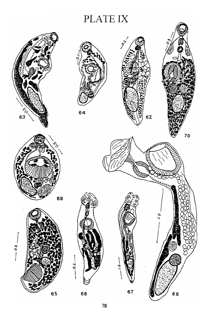
Figs. 66, 67 Plate IX
HOSTS:
SITE:
DATE OF
COLLECTION :
DEPOSITED
SPECIMEN :
Liza vaigiensis (Mugilidae) 1 11 of 8
Mugil cephalus (Mugilidae) 111 of 9
Intestine
Jan. 19, 1992,
Jan. 3 1 , 1992
USNPC 83986
Description and measurements based on two specimens, one mature from L.
vaigiensis, the other immature from M. cephalus , (measurements on immature
specimen in parentheses) . Body elongate, rounded posteriorly, 3 .380 (0.90)0 long ,
0. 875 (0. 350) wide at acetabular level; forebody 0.400 long. Cuticle aspinose, eye-spot
pigments very few , near pharynx. Oral sucker replaced by a broad membranous
muscular fold with six short, rounded lobes, as described by Manter (1961); ventral
sucker cup-shaped 0. 600 (0. 240) long , 0. 540 (0. 240) wide. Prepharynx short 0. 125
(0.095) long, 0.075 (0. 020) wide; pharynx ovoid , 0.350 (0. 125) long , 0. 250 (0. 105)
wide; esophagus short; cecum extending to anterior level of ovary . Testis elongate,
0. 675 (0. 105) long , 0. 275 (0.080) wide, near posterior end of body ; sperm duct
tubular, straight, becoming a seminal vesicle just posterior to the ventral sucker;
prostatic duct ovoid, dorsal to ventral sucker, with few prostatic cells in its anterior
half. Cirrus sac, if present, poorly developed. Ovary entire, overlapping anterior end
of testis, 0. 180 long, 0. 170 wide; seminal receptacle globular, thick walled, little
smaller than ovary; uterus not extending posterior to testis; eggs large, opaque ones 80-
100 long, 50-63 wide; vitellaria consisting of fused follicles, extending from posterior
end of testis laterally, but not reaching ventral sucker. Terminal parts of male and
female reproductive structures not discerned clearly anterior to mid acetabular level .
This is the sixth report of this species. Manter (1961) described it from an
unknown mullet (family Mugilidae) from Fiji. Durio and Manter (1968) reported it
from New Caledonia from an unknown mullet (family Mugilidae) and from M.
cephalus from Heron Island, Queensland , Australia. Hafeezullah (1971) found it in
Chirocentrus dorab from India. Overstreet in Lester and Sewell (1989) recorded it
from Crenimugil labiosus, L. vaigiensis, and Myxus elongatus. Cribb et al. (1994)
found it in Crenimugil crenilabis, M. cephalus, and L. vaigiensis from the southern
Great Barrier Reef, Australia. Our mature specimen is in general agreement with
Manter's description but differs in having a larger and more elongate testis. The testis
also occupies a more posterior location. Cribb et al. (1994), studying serial sections,
concluded that H. mulli possesses a vesicular pars prostatica but lacks a cirrus sac. We
have reexamined our specimen but are unable to see clearly any structure anterior to
the ventral sucker. We have also examined a paratype from Manter' s collection
(HWML 0510-F4) and a specimen from the New Caledonia (1847); the paratype shows
a membrane near the anterior level of the ventral sucker which could represent the
24

4 Pages 31-40

▲back to top

4.1 Page 31

▲back to top
lining of a cirrus sac or that of a vesicular pars prostatica as described by Cribb et al .
( 1 994) ; In the New Caledonia specimen a thin membrane is also seen which we
interpret as suggestive of a weakly developed cirrus sac thus confinning a conclusion
arrived at by Overstreet ( 1 969) in his examination of the same New Caledonia material ;
a short cirrus extruding from the genital pore was also seen ; the testis, though not as
elongate as the Fijian' s specimen, also occupies a more posterior position. It should be
noted , however, that these observations are based on whole mounts and subject to
personal interpretations; only Cribbs' specimens are based on sectioned material. Our
own conclusion is that a weakly developed cirrus sac is present.
The next group of haplosplanchnids have been reported from other parts of the
world and are presented as a review and an update.
PART II - Haplosplanchnids of Other Parts of the World
7. Haplosplanchnus mugilis Nahhas and Cable, 1 964
Fig . 30 Plate VI
This species was described from 13 specimens taken from Cur�ao, West
Indies.
According to Nahhas and Cable ( 1 964: 1 80)
"this i s the first species of Haplosplanchnus to be
reported from the Gulf-Caribbean region. Of the 3
previously described species H. mugilis is most like H.
pachysomus (Eysenhardt, 1829) , but differs in having a
shorter seminal vesicle, shorter peduncle of the ventral
sucker, a spherical or ovoid pars prostatica and the
uterus extending posterior to the testis. It differs from H.
caudatus Srivastava, 1 939 in the same respects and also
in body shape. In H. purii Srivastava, 1 939, the testis is
in the extreme posterior of the body , the cecum and
seminal vesicle are long, the vitellaria are compact, and
the pars prostatica tubular. "
Bray ( 1 984: 280) suggested that the extent of the uterus posterior to the testis
"may be of little importance and, in geographical terms, an intermediate form may
ocuc r. " We agree, in principle, with Bray ' s comments. We note, however, that none
of the species he placed in synonymy with H. purii, none of the Fijian specimens, or
H. otolithi which we also consider a synonym, have uterine coils that extend posterior
to the testis. Madhavi's (1979) observations on H. caudatum dealt with variations in
body shape and length of cecum but not on extent of the uterus. She did not report any
variations in extent of uterus in her specimens of H. purii either.
25

4.2 Page 32

▲back to top
HOSTS :
RECORD S :
GEOGRAPHIC
DISTRIBUTION:
Che/on labrosus, Crenimugil crenilabis, Mugil cephalus, M.
curema (Type host) , M. saliens, M. trichodon (Mugilidae).
Skinner ( 1975); Fischthal ( 1977). Juarez-Arroyo and Salgado­
Maldonado ( 1989). According to Bray ( 1984) , H. mugilis has
been reported, but not described, from the Mediterranean by
Nikolaeva and Parukhin in 1969, and by Parukhin, Naidenova
and Nikolaeva in 197 1 .
Curacao, Bay of Biscayne , Belize, Mediterranean, Mexican
Pacific
8 . Haplosplanchnus venezuelensis Fischthal and Nasir, 1974
Fig. 35 Plate VI
H. venezuelensis was described from a single specimen.
Fischthal and Nasir ( 1974: 73) stated that this species
" . . . appears closest to H. mugilis Nahhas and Cable,
1964 from a mugiliform (Mugilidae) fish from Curacao.
The latter species differs from H. v�lensis in having
the testis about midway between the ventral sucker and
posterior extremity and the genital pore midway between
the ventral sucker and pharynx, and the vitellaria
consisting of 10-12 inconspicuous, scattered follicles. "
HOST:
RECORD :
GEOGRAPHIC
DISTRIBUTION :
An tennarius multiocella tus (Antennariidae).
Fischthal and Nasir ( 1 974)
Los Roques Islands (Venezuela)
9. Haplosplanchnus bengalensis Gupta and Puri , 1 985
Fig. 37 Plate VI
H. bengalensis was desc ribed from a single specimen .
In comparing this species to others Gupta and Puri ( 1985 : 56) indicated that
" . . . the new form has close resemblance with H. purl and
H. indica in having genital papillae lying on prominent
conical papillae but differs from them in having
intestinal caeca extending up to a little posterior to
ventral sucker instead of up to a little anterior to ovary
in H. purii and up to anterior third of body in H. indica.
The new form further differs from H. purii in the extent
of the uterus between anterior margin of testis and
oesophagus instead of from vitellaria to pars prostatica.
26

4.3 Page 33

▲back to top
Accordingly it is regarded as a new species with the
specific name Haplosplanchnus bengalensis sp. nov. "
This species, has a strong resemblance, except for more extensive vitellaria, to
H. elongatus and H. indicus both of which were considered synonyms of H. purii by
Bray ( 1 984) . The vitellaria were described by Gupta and Puri ( 1 985 : 54) as
"follicular, poorly developed, extending from anterior
end of pharynx up to a little anterior to hind end of body
but interrupted in oesophageal region. "
This distribution of vitellaria is unusual for the genus Haplosplanchnus and
needs confirmation.
HOST:
RECOR D:
GEOGR APHIC
DISTRIBUTION :
Acanthurus sandvicensis ( Acanthuridae)
Gupta and Puri ( 1 985) .
Puri, Orissa, Bay of Bengal.
10. Haplosplanchnus vinodae Ahmad, 1 985
Fig. 38 Plate VI
H. vinodae was described from seven specimens.
Ahmad (1985: 191) distinguished it
"from all the known species of the genus
Haplosplanchnus in having prostate vesicle bipartite
instead of unipartite. "
In many respects this species is very similar to H. otolithi except for its
'bipartite' prostate vesicle and the extent of the uterus to the posterior end of the body.
Ahmad's figure shows what looks to us like a ' split' rather than a ' bipartite' prostatic
vesicle.
HOST:
RECOR D:
GEOGRAPHIC
DISTRIBUTION:
Mugil cephalus
Ahmad ( 1 985)
Panjim coast, Goa, India.
27

4.4 Page 34

▲back to top
1 1 . Schikhobalotrema acutum (Linton, 1910) Skrj . and Gusch. , 1 955
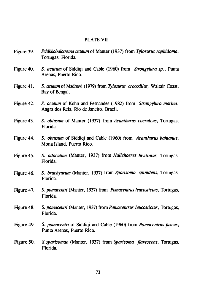
Figs. 39-42 Plate VII
SYNONYMS : Distomum spp. Linton, 1 907
Deradena acuta Linton, 1910
Haplosplanchnus acuta (Linton, 1910) Manter, 1937
Linton's (1910) original description is based on specimens recovered from the
intestine of Tylosurus acus from Bermuda and T. marinus from Tortugas, Florida.
Manter (1937: 386) redescribed the species from several specimens from T. raphidoma
from Tortugas, stating
"This species differs from all preceding species of
Haplosp'lanchnus in the longitudinal slit-like aperture of
the ventral sucker and the pair of flap-like lateral
processes at its posterior end . These characters may
prove to be of generic value. The genital bulb is much
better developed in H. acutus and the eggs are slightly
smaller than in preceding species from Tortugas.
What seme s to be the same species had bene collected by
the author from Tylosurusfodiator and Kyphosus elegans
from the Pacific Ocean at Port Utria, Colombia. It may
prove to be a characteristic and widely distributed
parasite of Tylosurus. The lobe-like processes of the
ventral sucker are unique. They were apparently
overlooked by Linton and no observations on their living
state are available. They seem to be flexible, perhaps
contractile. Since they do not seem to be strong enough
to aid in attachment they may serve as sense organs" .
Overstreet (1969) described the tubular genital bulb. Madhavi ( 1 979:240) stated
that her 24 specimens are:
" . . . morphologically indistinguishable from those
described from American Atlantic and Pacific
Coasts. . However, a notable feature in the present
specimens is that they lacked spermatozoa inside the
reproductive ducts. In inunature as well as gravid
specimens the receptaculum seminis and seminal vesicle
has a collapsed appearance and they are filled with a
granular material rather than spermatozoa. Earlier
authors illustrated receptaculum seminis and seminal
vesicle of S. acutum as being distended with
spermatozoa. To confirm the absence of spermatozoa in
the present specimens section and smears of testis stained
28

4.5 Page 35

▲back to top
with Feulgen were examined. They revealed but few
spennatozoa which appear abortive. The eggs produced
by these flukes, however, developed nonnally. It appears
that this is a parthenogenic race and detailed cytological
study of this fonn may prove that it is a sibling species. 11
HOSTS :
RECORDS:
GEOGRAPHIC
DISTRIBUTION:
Thyrinops pachylepis (Atherinidae); Strongylura ardeola, S.
timucu, Strongylura sp., Tylosurus acus, T. caribbaeus, T.
crocodilus, T. marinus, T. raphidoma (Belonidae);
Hemiramphus margintatus H. georgi, Hyporamphus unifasciatus
(Hemiramphidae); Mugil cephalus (Mugilidae); Abudefduf
sartilis (Pomacentridae) ; Kyphosus elegans(Kyphosidae) ;
Lachnolaimus maximus (Labridae).
Linton (1910); Manter (1937 , 1940 , 1 947); Caballero et al.
(1 95 3); Perez Vigueras (1956) ; Siddiqi and Cable (1 960);
Sogandares-Bemal and Sogandares (1 961) ; Nahhas and Cable
(1 964); Overstreet (1 969) ; Fischthal (1977); Madhavi (1 979);
Yeo and Spieler (1 980) , Kohn and Fernandes (1 982 ; Fayek et
'
al. (1 990) ; Shen (1 990)
Bermuda, Florida, Brazil, Galapagos Islands, Pacific coast of
Colombia, Cuba, Puerto Rico, Curacao, Jamaica, Belize, Bay
of Bengal, Mediterranean, South China Sea.
12. Schikhobalotrema obtusum (Linton, 1 910) Skrj . and Gusch. , 1 955
Figs. 43 , 44 Plate VII
SYNONYMS: Deradena obtusa Linton, 1 910
Haplosplanchnus obtusus (Linton, 1 910) Manter, 1 937
Manter (1 937 : 38 2) distinguished this species
from 11 • • • all others at Tortugas by the almost equal size
of the suckers and the elongated seminal receptacle. The
hindbody is long, the ovary unlobed in contrast to certain
other species. It differs from H. pachysomus in length of
cecum, in extent of v!tellaria and in larger size of eggs. "
Pritchard and Manter's record from Hawaii was questioned by Yamaguti (1 971)
who indicated that the Hawaiian form is probably S. acanthuri Yamaguti , 1 971 .
29

4.6 Page 36

▲back to top
HOSTS:
RECORDS :
GEOGRAPHIC
DISTRIBUTION:
Acanthurus achilles, A. bahianus, A. caerulus, A. sandvicensis,
Paracanthurus hepatus ,( = Acanthurus hepatus)(Acanthuridae) ,
Sparisoma viride (Sparidae) .
Linton (1910); Manter ( 1937 , 1947); Siddiqi and Cable ( 1 960);
Nahhas and Cable ( 1 964); Pritchard and Manter ( 1 96 1 ); Nahhas
and Carlson ( 1994).
Florida, Puerto Rico , Curacao , Jamaica, Hawaii .
1 3 . Schikhobalotrema adacutum (Manter, 1 937) Skrj . and Gusch. , 1 955
Fig. 45 Plate VII
SYNONYM : Haplosplanchnus adacutus Manter, 1937
According to Manter ( 1937: 387)
" it is very difficult to separate this species from
Haplosplanchnus acutus . It is distinctly smaller, only
one specimen of over 20 reaching a length of 1 mm,
whereas the smallest of 6 specimens of H. acutus was
1 . 267 mm. In all except one specimen of H. adacutus,
the posttesticular distance was greater than the testis­
ventral sucker distance, whereas the opposite was true in
all specimens of H. acutus. The ventral sucker lobes
were almost always very inconspicuous in H. adacutus
(except two specimens from Halichoeres maculipinnus)
and very evident in H. acutus. A constant difference
occurs in the relative size of the genital atrium or bulb
which is small and inconspicuous in H. adacutus and as
large or larger than the pharynx in H. acutus. These
differences may be those of one species in different hosts
but the general impression given is that the two forms
(each of which was collected several times) are distinct . "
HOSTS :
RECORDS :
GEOGRAPHIC
DISTRIBUTION:
Abudefduf marginatus, A. saxatilis (Pomacentridae);
Halichoerus maculipinnus , H. bivittatus, H. pictus (Labridae);
Hemiramphus brasiliensis (Hemiramphidae); Hepsetia stipes
(Atherinidae) .
Manter ( 1 937, 1947); Nahhas and Cable ( 1 964) , Yeo and
Spieler ( 1 980) .
Florida, Jamaica, Curacao .
30

4.7 Page 37

▲back to top
14. Schikhobalotrema brachyurum (Manter, 1937) Skrj . and Gusch. , 1955
Fig. 46 Plate VII
SYNONYM: Haplosplanchnus brachyurus Manter, 1937
According to Manter ( 1961 : 384)
"this species differs from the others at Tortugas by the
short hindbody , large ventral sucker and anterior extent
of vitellaria. In these respects it is especially distinct
from H. obtusus. The sucker ratio is approximately that
of H. sparisomae but the hindbody is much shorter and
the ovary is unlobed. "
HOSTS:
RECORDS:
GEOGRAPHIC
DISTRIBUTION:
Cryptotomus auropunctatus, PseTJ(foscarus guacamaia,
P. coelestinus (Scaridae); Sparisoma aurofrenatum, S.
spinidens, S. viride (Sparidae).
Manter ( 1937, 1947).
Florida.
15. Schikhobalotrema pomacentri (Manter, 1937) Skrj. and Gusch. , 1955
Figs. 47-49 Plate VII
SYNONYM : Haplosplanchnus pomacentri Manter, 1937
According to Manter ( 1 937:385)
"this species was not reported by Linton. It differs from
all others at Tortugas in the presence of a distinct
prostatic vesicle with tall transparent cells and in a more
muscular genital atrium. The species most similar to it is
H. brachyurus from parrot fish. But in addition to the
point noted, H. pomacentri differs in that the hindbody
is never shorter that the forebody . "
Manter ( 1 937: 385) also indicated that
" . . . a Haplosplanchnus species from Pomacentrus
rectifraenum Gill collected in the Galapagos Islands
agrees with H. pomacentri from Tortugas in all
important respects including the prostatic vesicle and is
probably the same species. "
31

4.8 Page 38

▲back to top
Nahhas and Carlson ( 1 994) listed its most important characteristics: a relatively
smal l seminal receptacle, almost equal length of fore- and hindbody , smal l ovary
(about same size as egg length) , transverse acetabular aperture, tendency of vitellaria
to coalesce longitudinally, muscular genital atrium, and a distinct prostatic vesicle.
Cribb et. al ( 1 994) reported this species from several pomacentrids; their figure shows
a ventral sucker slightly anterior to midbody but more importantly eggs much smaller
than the ovary ; the measurements they give in their Table II do not include egg size for
comparison with ovary size; In other respects, it has many similarities to S.
pomacentri. Based on their figure, their specimens do not key out to S. pomacentri or
any known Schikhobalotrema species (no key is really adequate or good enough to
make a definitive identification especially if based on a drawing) . The Australian
species may represent a new form but a comparison between ovary size and egg length
should be made before definitive diagnosis is made; for the time being we accept the
authors' identification. S. pomacentri was not found in 1 3 fishes belonging to six
species of pomacentrids from Fij i (unpublished data) .
Fischthal' s (1977) report of S. pomacentri from Sparisoma chrysopterus is the
only report of this species from a non-pomacentrid host.
HOSTS :
RECORDS :
GEOGRAPHIC
DISTRIBUTION:
Abudefduf whitleyi, Acanthochromis polyacanthus, Amphiprion
akinodynos, A. perideraion; Microspathodon chrysurus;
Panna polylepis; Chromis chrysura ( = Pomacentrus chrysurus),
C. Xanthura ( = Pomacentrus xanthurus), C. analis
(Pomacentrus analis), Pomacentrus fuscus, P. leucostictus, P.
philippinus, P. planifrons, P. rectifraenum, P. tripunctatus,
Pomacentrus sp., Stegastes apicalis (Pomacentridae) ; Sparisoma
chrysopterum (Sparidae).
Manter ( 1937, 1 947); Siddiqi and Cable ( 1960); Sogandares­
Bernal and Sogandares (1961); Nahah s and Cable (1964) ;
Fischthal ( 1 977) ; Yeo and Spieler (1980); Dyer et a/. (1991);
Nahah s and Carlson (1994) ; Cribb et al. (1 994).
Florida, Puerto Rico , Panamanian Atlantic , Jamaica, Belize,
southern Great Barrier Reef.
16. Schikhobalotrema sparisomae (Manter, 1937) Skrj . and Gusch. , 1955
Fig. 50 Plate VII
SYNONYM: Haplosplanchnus sparisomae Manter, 1937
Manter ( 1 937: 382) distinguished this species
" . . . from others at Tortugas by the large ventral
sucker with circular aperture and the deeply
lobed or tripartite ovary. It differs from H.
32

4.9 Page 39

▲back to top
pachysomus in sucker ratio, shape of ovary,
extent of vitellaria and in its much larger eggs. "
Nahhas and Carlson (1994 :11) stated
"Posterior extent of the seminal vesicle was not
described by Manter (1937) nor mentioned by
other autho rs. One of the five specimens clearly
shows a long seminal vesicle which is almost
straight extending posteriorl y to level of ventral
sucker. A reexamination of th ree worms
identified as this species from Pseudoscarus
guacamaia from me earlier Jamaican collection
shows, in one worm, a seminal vesicle
extending only to mid- acetabular level. "
HOSTS:
Mugil cephalus (Mugilidae) ; Nicholsina usta, Pseudoscarus
guacamaia, P. perrico, Scarus croicensis, Scarus sp . (Scaridae) ;
. Sparisoma abildgaardi, S. brachiale, S. jlavescens, S. radians, S.
viride (Sparidae) .
R EC ORDS:
Manter (1937) ; Nahhas an d C able (1964) ;
Overstree t (1969) ; Nahhas and Carlson (1994).
GEOGRAPH IC
DISTRIBUTION: Florida, Jamaica, C ur�ao.
1 7 . Schikhobalotrema kyphosi (Manter, 1947) Skrj. an d Gusch. , 1955
Fig. 51 Plate VIII
SYNONYM: Haplosplanchnus kyphosi Manter, 1947
According to Manter (1947 :3 26)
" . . .H. sparisomae is the only other species with
lobed ovary and seem to be the most closely
related species. It differs in body shape , sucker
ratio (the ventral sucker is 1 .5 times the oral
sucker) , in extent of uterus, and in its very sh ort
posttesticular sp ace. "
HOSTS:
Kyphosus incisor, K. sectatrix (Kyphosidae).
REC ORDS:
Manter (1947) ; Overstree t (1969) .
GEOGRAPH IC
DISTRIBUTION : Florida (Tortugas , Bay of B iscayne)
33

4.10 Page 40

▲back to top
18. Schikhobalotrema girellae (Manter and van Cleave, 195 1 ) Skrj. and Gusch. ,
1 955
Fig. 52 Plate VIII
SYNONYM: Haplosplanchnus girellae Manter and Van Cleave, 1951
According to Manter and Van Cleave ( 1 95 1 : 335)
"H. girellae is probably most similar to H.
kyphosi Manter, 1947, and H. obtusus (Linton,
1 9 10) from Tortugas, Fla. It differs from H.
obtusus in its long posttesticular space, in the
spherical shape of the seminal receptacle, in
J acking distinct vitelline follicles, in the
interruption of the vitellaria opposite the testis,
and in shape of the eggs. It differs from H.
kyphosi in its unlobed ovary, relatively larger
ventral sucker, elongated testis, interrupted
vitellaria and smaller body sire. "
HOST:
Girella nigricans Kyphosidae
RECORDS:
Manter and Van Cleave ( 1951).
GEOGRAPHIC
DISTRIBUTION: La Jolla (California) .
19. Schikhobalotrema adbrachyurum Siddiqi and Cable, 1960
Fig. 53 Plate VIII
According to Siddiqi and Cable ( 1 960: 275)
"S. adbrachyura resembles most S. brachyura
but differs from it in having a strongly lobed
ovary and shorter seminal vesicle. "
HOSTS :
Pseudoscarus guacamaia, P. plumbaeus (Scaridae) ; Sparisoma
jlavescens, S. viride, Sparisoma sp. (Sparidae).
RECORDS:
Siddiqi and Cable ( 1 960); Nahhas and Cable (1 964)
GEOGRAPHIC
DISTRIBUTION: Puerto Rico, Jamaica, Curacao.
34

5 Pages 41-50

▲back to top

5.1 Page 41

▲back to top
20. Schikhobalotrema manteri Siddiqi and Cable, 1960
Fig . 54 Plate VIII
According to Siddiqi and Cable ( 1 960: 274)
"Schikhobalotrema manteri differs from all other
species of the genus in the distribution of
vitellaria, the relative position of the gonads,
and the posterior extent of the uterus. "
HOSTS :
a "reeffish with a collar "; Acanthurus bahianus (Acanthuridae)
RECORDS:
Siddiqi and Cable ( 1 960); Dyer et al. ( 1 992) .
GEOGRAPHIC
DISTRIBUTION: Puerto Rico.
21 . Schikhobalotrema crasswn Pritchard and Manter, 1 96 1
Fig . 5 5 Plate VIII
According to Pritchard and Manter ( 1 96 1 : 195)
"S. brachyurum and S. hawaiensis may also
have the testis near or even overlapping the
ventral sucker. S. brachyurum has conspicuously
larger suckers relative to body siz.e, although the
sucker ratio is probably similar, the hindbody is
much shorter, and the vitellaria are more
extensive and more follicular. S. hawaiensis has
a shorter hindbody , a smaller sucker ratio, lacks
large prostatic cells, lacks a well-differentiated
metraterm, and lacks the conspicuously thick
cuticula. "
Pritchard and Manter ( 1 96 1 : 196) noted that
" . . . S. crassum also resembles S. robustum with
which it may be sympatric but S. crassum has a
thicker body with a conspicuously thick cuticula,
the testis overlaps the ventral sucker 113 or more
with the ovary correspondingly more anterior,
and the excretory vesicle is shorter. "
HOST:
Stegastes fasciolotus ( =Pomacentrus jenkinsi) (Pomacentridae)
RECORD :
Pritchard and Manter ( 1 96 1).
GEOGRAPHIC
DISTRIBUTION : Hawaii
35

5.2 Page 42

▲back to top
22. Schikhobalotrema glomerosum Pritchard and Manter, 1961
Fig. 56 Plate VIII
According to Pritchard and Manter ( 196 1 : 196)
" . . . S. brachyurum (Manter, 1 937) and S.
adbrachyurum Siddiqi and Cable, 1960 are the
only other species in which the ventral sucker
may be found posterior to the midbody . In both
species the cecum extends posterior to the
ventral sucker, the ventral sucker is larger, the
ovary and testis are in the hindbody, the testis is
smaller than the ventral sucker, the vitellaria are
follicular, and the body is more elongate. "
HOSTS:
Acanthurus achilles, A. sandvicensis (Acanthuridae).
RECORD:
Pritchard and Manter ( 1 961).
GEOGRAPHIC
DISTRIBUTION: Hawaii.
23. Schikhobalotrema hawaiiensis Pritchard and Manter, 1961
Fig. 57 Plate VIII
According to Pritchard and Manter ( 1 96 1 : 194)
"three species, S. brachyurum (Manter, 1 937),
S. pomacentri (Manter, 1937), and S.
adbrachyurum Siddiqi and Cable, 1960, have
the ventral sucker located at or slightly behind
the midbody . Tortugas, Florida, is the type
locality for the first two species and Puerto Rico
is the type locality for S. adbrachyurum. Manter
(1940) has also reported S. pomacentri from the
Galapagos Islands. " S. hawaiensis is like S.
pomacentri in that the hindbody may equal the
forebody in length but is never shorter, and the
anterior vitellaria do not extend to the oral
sucker. S. hawaiensis, however, differs from S.
pomacentri by lacking a prostatic vesicle, by
lacking a posterior marginal projection on the
oral sucker, by a less tapered and more rounded
hindbody , in the testis which is larger than the
ventral sucker and immediately posterior to it
(rather than being in the middle of the
36

5.3 Page 43

▲back to top
hindbody). S. hawaiensis is like S. brachyurum
in lacking the prostatic vesicle, in lacking a
posterior marginal projection of the oral sucker,
in the testis that may overlap the ventral sucker,
and the seminal vesicle that is sinuous rather
than almost straight. It differs from S.
brachyurum in that the hindbody is never shorter
than the forebody (though it may be nearly
equal), the vitellaria usually extend only to the
level of the genital pore (and no farther forward
than the posterior edge of the pharynx), the
uterus is entirely preovarian, and the ventral
sucker is slightly smaller affecting the sucker
ratio (1: 1. 2 to 1. 4 as compared with 1: 1. 5 to 2
for S. brachyurum) . S. adbrachyurum has the
ventral sucker slightly posterior to midbody, but
the testis is near the posterior end of the body,
the ovary is lobed, the seminal vesicle is shorter,
the eggs are wider, and the body is smaller"
Yamaguti (1970) indicated that a metraterm is present, contrary to Pritchard and
Manter' s (1961) assertion of its absence. Yamaguti 's specimens were obtained from
the same host species and locality as Pritchard and Manter' s.
HOSTS :
Acanthurus mata, CtetWchaetus strigosus (Acanthuridae) .
RECORDS:
Pritchard and Manter (1961) ; Yamaguti (1970) .
GEOGRAPHIC
DISTRIBUTION : Hawaii .
24. Schikhobalotrema robustum, Pritchard and Manter, 1961
Fig. 58 Plate VIII
According to Pritchard and Manter ( 1 96 1 : 192)
" in size and long posttesticular space S.
robustum is similar to S. kyphosi (Manter,
1947), but it differs markedly in having an
unlobed ovary, much larger testis, larger sucker
ratio, and less follicular vitellaria. S. robustum
i s most similar to S. girellae (Manter and Van
Cleave, 195 1) . It differs in being much larger,
although the egg size is about the same. The
difference in body size makes the eggs appear to
be much larger in S. girellae. The so-called
cirrus of S. girellae is probably a thick-walled
tubular atrium although entrance of the uterus
37

5.4 Page 44

▲back to top
could not be sene in 15 paratypes. The atrial
tube in S. robustum is thin-walled and shorter.
Prostatic cells are much smaller in S. girellae.
Although the interruption of the vitelline glands
opposite the testis was emphasiz.ed for S.
girellae, we now find that indistinct strands of
vitelline material connect the pre- and
posttesticular vitelline masses in some of the
paratype specimens. In S. robustum the dorsal
connections are conspicuous" .
HOSTS:
Acanthurus sandvicensis, Zebrasoma flavescens (Acanthuridae);
Chaetodon fremblii (Chaetodontidae); Stegastes fasciolatus
( = Pomacentrus jenkinsi) (Pomacentridae) .
RECORDS :
Pritchard and Manter (1961); Yamaguti (1970) ; Juarez-Arroyo and
Salgado-Maldonado (1989).
GEOGRAPHIC
DISTRIBUTION: Hawaii, Mexican Pacific
25 . Schikhobalotrema bivesiculum Nahhas and Cable, 1964
Fig. 59 Plate VIII
According to Nahhas and Cable ( 1964: 182)
"Schikhobalotrema bivesiculum differs from all
other species of the genus in its conspicuous
bipartite seminal vesicle. It resembles S. acutum
and S. adacutum in general topography and
although it has a longitudinal aperture in the
ventral sucker as in those species, the sucker
lacks posterolateral lobes. The bipartite seminal
vesicle with a thick anterior division in this
species may be of generic value but the species
is placed in Schikhobalotrema because others
showing gradations in that respect may exist. "
S. bivesiculum 's original description was based on six specimens from A.
saxatilis from the south shore of Jamaica. Nine specimens of this species from the same
host species were also reported by Nahhas and Carlson (1994:9) from the north shore
of Jamaica who added the following observations:
"the muscular (anterior thick part) of the seminal
vesicle is one to one and one-half length of the
pharynx and may be globular or cylindrical in
shape; a shon prostate duct is present but no
38

5.5 Page 45

▲back to top
prostate cells are seen; the genital atrium is
conspicuous but not muscular; the ovary is
usually anterior to testis but is dorsal to it in one
specimen and dorsal to the ventral sucker in
another. In the live material the excretory
vesicle seemed to reach the testis, level
depending on degree of contraction. The
aperture of the ventral sucker is a narrow
longitudinal slit in some but broadly so in
others. "
HOST:
Abudefduf saxatilis (Pomacentridae).
RECORDS :
Nahhas and Cable (1964); Nahhas and Carlson ( 1 994) .
GEOGRAPHIC
DISTRIBUTION: Jamaica.
26. Sthikhobalotrema elongatum Nahhas and Cable, 1964
Fig. 60 Plate VIII
According to Nahhas and Cable ( 1 964: 182)
" t he
most
distinctive
features
of
Schikhobalotrema elongatum are its long
hindbody with prominent cuticular rings, and its
well-developed bulb of prostatic ducts. In body
shape, S. elongatum is similar to several species.
S. acutum and S. adacutum have a ventral
sucker with a longitudinal aperture and lateral
lobes. S. obtusum lacks a prostatic vesicle; the
genital atrium is non-muscular and the ovary
close to the ventral sucker. S. pomacentri has an
equat0rial ventral sucker and S. kyphosi
(Manter, 1947) a lobed ovary. S. girellae
(Manter and Van Cleave, 1 95 1) is most like S.
elongatum but differs from that species in the
extent of the seminal vesicle, in having a long
tubular genital atrium and a more anterior
ovary. S. manteri Siddiqi and Cable, 1960,
differs in the distribution of the vitellaria,
position of the testis and posterior extent of the
uterus. S. robustum Pritchard and Manter, 1961 ,
is much larger , has a thin-walled genital atrium
and the genital pore on a finger-like projection. "
39

5.6 Page 46

▲back to top
HOSTS:
Mugil cephalus, M. curema (Mugilidae).
RECORDS:
Nahhas and Cable ( 1 964); Skinner ( 1 975).
GEOGRAPHIC
DISTRIBUTION: Jamaica, Florida.
27. Schikhobalotrema acanthuri Yamaguti, 1970
Fig. 61 Plate VIII
According to Yamaguti ( 1 970: 25)
"this species resembles Schikhobalotrema
obtusum (Linton, 19 10) , but differs from it in
the ovary being far posterior to the ventral
sucker and in the uterus not extending posterior
to the posterior end of the seminal vesicle which
reaches to the ovary. S. obtusum (Linton, 1910),
found by Pritchard and Manter ( 1 961) in
Acanthuros sandvicensis and other Acanthurus
species of Hawaii, probably belongs to the
present species. "
HOSTS:
Acanthurus sandvicensis, A. nigrofascus, Zebrasoma flavescens
(Acanthuridae) , Mugil liza (Mugilidae)
RECORDS :
Yamaguti (1970) , Fernandes and Goulart ( 1989)
GEOGRAPHIC
DISTRIBUTION : Hawaii; Praia de Copacabana, Rio de Janeiro State (Brazil)
28. Schikhobalotrema longivesiculatum Orecchia and Paggi , 1975
Fig. 62 Plate IX
According to Orecchia and Paggi ( 1 975 : 74), whose description is based on 1 1
specimens ,
"Schikhobalotrema longivesiculatum differs
from Schikhobalotrema obtusa (Linton, 1910)
Skrjabin and Guschanskaja, 1955, especially for
sucker ratio, lobed ovary, seminal vesicle coiled
and in larger size of eggs; from
SchikhobalotrelTUl pomacentri (Manter, 1937)
Skrjabin and Guschanskaja, 1955 for lobed
ovary, elongated seminal receptacle, smaller
vitelline follicles and coiled seminal vesicle;
from Schikhobalotrema sparisomae (Manter,
1 937) Skrjabin and Guschanskaja, 1955 for
40

5.7 Page 47

▲back to top
elongated seminal receptacle, coiled seminal
vesicle and extent of excretory vesicle. "
HOST:
Blennius sanguinolentus (Blenniidae)
RECORDS :
Orecchia and Paggi ( 1 975) .
GEOGRAPHIC
DISTRIBUTION: Gulf of Gaeta (Italy) .
29. Schkhobalotrema magnum Sknner, 1 975
Fig. 63 Plate IX
According to Skinner (1975: 328)
"the most distinctive features of S. magnum are
its large siz.e, comparatively small oral sucker,
ventral sucker, and eggs, extensive vitellaria,
large testis and lobed ovary, and the structure of
its terminal genitalia. S. magnum is similar to S.
.
kyphosi (Manter,
Tortugas, and S.
1 947) from Kyphosus sp. ,
sparisomae (Manter, 1937)
from Sparisoma sp. , also from Tortugas, in its
lobed ovary and large siz.e. S. magnum differs
from S. kyphosi in its larger testis, larger sucker
ratio, and less follicular vitellaria. It differs
from S. sparisomae in its smaller sucker ratio,
a shon cecum, sinuous seminal vesicle. and the
size of the eggs. In overall siz.e and proportions
S. magnum is most like S. robustum Pritchard
and Manter, 1 96 1 , from Hawaiian fishes. The
extension of the vitellaria, large siz.e, sinuous
seminal vesicle with a distal constriction, and
the presence of large prostatic cells are similar
in both species. S. robustum differs mainly in
having its genital pore on a finger-like
projection, the lack of a prostatic vesicle, and an
unlobed ovary. "
HOST:
Mugil cephalus (Mugilidae)
RECORD:
Skinner (1 975) .
GEOGRAPHIC
DISTRIBUTION: Biscayne Bay (Florida) .
41

5.8 Page 48

▲back to top
30. Schikhobalotrema solitaria Fernandes and Goulart, 1989
Fig 71 Plate X
According to Fernandes and Goulart (1989) who compared the single specimen
of their new species to the 19 species of Schikhobalotrema the most distinctive feature
of S. solitaria is the ratio of the oral sucker to the pharynx which is 1 : 1 . 1 whereas in
all the others it is 1 : 0. 3-0. 6.
HOST:
RECORDS :
GEOGRAPHIC
DISTRIBUTION:
Stephanolepis hispidus (Balistidae)
Fernandes and Goulart ( 1 989)
Praia de Copacabana, Rio de Janeiro State (Brazil)
3 1 . Pseudoschikhobalotrema heterocotylum (Nahsah ans Cable, 1964),
Yamaguti, 197 1
Fig. 69 Plate IX
SYNONYM: Sckikhobalotrema heterocotylum Nahhas and Cable, 1964
According to Nahhas and Cable ( 1 964 : 13)
"no haplosplanchnid has bene described as
having a ventral sucker with the tuberculated
lobes characteristic of this species. Most like it
in other respects is S. brachyurum (Manter,
1937) a paratype of which we have examined
and found to lack such lobes. It further differs
from S. heterocotylwn in having a different body
shape; S. glomerosum Pritchard and Manter,
1 961 , has a smaller ventral sucker, a
preacetabular ovary, more anterior testis and a
well-developed metraterm. "
P. heterocotylum was originally described from P. guacamaia from Cur�ao.
Four specimens were reported from S. croicensis from the nonh shore of Jamaica by
Nahhas and Carlson ( 1 994: 1 1) who stated
"The four large worms have an almost spherical
shape when studied alive. One was immature
and whitish in color, the other three were
mature with a brown-red coloration. All four
clearly showed the characteristic tuberculated
papillae or lobes of the ventral sucker
characteristic of this species.
42

5.9 Page 49

▲back to top
HOSTS:
Pseudoscarus guacamaia, Scarus croicensis (Scaridae).
RECORDS :
Nahhas and Cable ( 1964) ; Nahhas and Carlson ( 1994) .
GEOGRAPHIC
DISTRIBUTION: Cur�ao, Jamaica.
32. Haplosplanchnoides hemiramphi Nahhas and Cable, 1964
Fig. 65 Plate IX
According to Nahhas and Cable ( 1 964: 183)
"although certain other haplosplanchnids have
short hindbodies, their gonads are never anterior
to the ventral sucker except perhaps in
Schikhobalotrema glomerosum which Pritchard
and Manter ( 1961) described from two
specimens in poor condition. "
HOST:
Hemiramphus brasiliensis (Hemiramphidae)
RECORDS : . Nahhas and Cable ( 1 964) .
GEOGRAPHIC
DISTRIBUTION: Jamaica
33. Hymenocotta manteri Overstreet, 1969
Fig . 68 Plate IX
SYNONYM: Hymenocottoides manten· (Overstreet, 1969) Yamaguti , 197 1
According to Overstreet ( 1969: 133)
"Manter ( 1 96 1 : 67-69) erected the genus
Hymenocotta to accommodate a single species,
H. mulli Manter, 196 1 , which differs from all
other haplosplanchnids in having a cirrus sac
and a six-lobed disc, which replaces the oral
sucker. The present species is the second with a
disc , but it is not six-lobed, and a cirrus sac is
absent. What appears to be the cirrus sac is
weakly developed and not evident in all of Dr.
Manter's specimens of H. mulli from New
Caledonia. Pending further infonnation� the
present species is placed in Hymenocotta
because of the oral disc which, with the absence
of a well-developed bulb of prostatic duct,
distinguishes H. monteri from Schikhobalotrema
43

5.10 Page 50

▲back to top
elongatum Nahhas and Cable, 1964 and
otherwise similar species from the same host. "
Yamaguti ( 1 971) removed H. monteri from the genus Hymenocotta and erected
a new genus Hymenocottoides, based on two distinguishing characteristics of H.
manteri: absence of a cirrus sac and presence of a bulbous prostatic vesicle free in the
parenchyma.
The original description of this species was based on 16 whole mounts and three
sectioned specimens from M. cephalus, Biscayne Bay, Florida; H. manteri was
collected again by Overstreet ( 1 97 1) from the same host species from artificial ponds,
Price Lake in Rockefeller Wildlife Refuge, Grand Chenier, Louisiana, and waters
adjacent to Mississippi Sound, Mississippi, redescribed it and found it to have a cirrus
sac .
According to Skinner ( 1 975) , H. manteri was also found in the same host
species by Rawson in 1973 along the coast of Georgia. Tantalean et al. ( 1982) and
Tantalean et al. ( 1992) also reported this species from the Peruvian coast.
Fernandez and Bendezu ( 1986) did not observe a cirrus sac in their specimens
of H. manteri from San Marcos, but Bariega ( 1 987) reported the presence of a weak
cirrus sac in specimens from M. cephalus in Chile.
HOST:
Mugil cephalus Mugilidae
RECORDS :
Overstreet ( 1969, 1 97 1 ) ; Rawson in Skinner
( 1 975) ; Fernandez and Bendezu ( 1 986) ; Bariega ( 1 987).
GEOGRAPHIC
DISTRIBUTION: Biscayne Bay (Florida), Louisiana, Mississippi, Georgia, San
Marcos, Chile, Peru.
34. Prohaplosplanchnus diorchis Tang and Lin, 1978
Fig. 20 Plate IV
Prohaplosplanchnus diorchis, described from several specimens, is the only
member of the family Haplosplanchnidae with two testes.
HOSTS :
Mugil cephalus, Liza haematochilus.
RECORD :
Tang and Lin ( 1 978)
GEOGRAPHIC
DISTRIBUTION: Min River, Fujian, China.
44

6 Pages 51-60

▲back to top

6.1 Page 51

▲back to top
35 . Provitellotrema crenimugilis Pan, 1984
Fig. 64 Plate IX
Pan ( 1984) erected the genus Provitellotrema, for P. crenimugilis based on a
single spec imen, placed it in the subfamily Sch ikhobalotremat inae , compared it with
Schikhobalotrema not ing se veral spec ific differences be tween the two genera:
" the vitellar ia of Schikhobalotrema is well­
developed, located at po ster ior end and sides of
body, whereas the v itellar ia of Provitellotrema
is div ided into two groups form ing an arch
between the phary nx and ventral sucker. The
ovary and testis of Schikhobalotrema are situated
at m idbody level; i n Provitellotrema the ovary
and testis are near po ster ior end of body. "
HOST:
Crenimugil crenilabis (M ugi l idae)
RECORD :
Pan (1984)
GEOGRAPHIC
DISTRIBUTION : C h ina.
36 . Neohaplosplanchnus ablennis Abdu l-Sal am and Khal il, 1987
Fig.70 Plate IX
According to Abd ul- Sal am and Khal il (1987:153) whose descr iption and
measurements were base d "on7 whole- mounts, 3 sets of sec t ioned speci mens and3
spec imens examined under the scanning elec tron m icrosc ope. "
"as the present spec imens cannot be a ssigned to
any of the ex isting genera of the fam ily
(Haplosplanchni dae) , a new genus is establi shed
for them. From Hymenocotta Manter 196 1 and
Hymenocottoides Yamagut i, 197 1 it differs i n
the presence of an oral sucker of the normal type
as opposed to a l obed disc. Fro m
Haplosplanchnoides N ahhas and C able, 1964 it
differs in the pre-equ ator ial po sit ion of the
ventral sucker instead of bei ng near the po sterior
extremity and in havi ng a shorter cec um not
reach ing the po steri or extremity. From
Haplosplanchnus Loo ss, 1902 and Laruea
Srivastava, 1939 , it differ s in hav ing an
extensive follicular vitel larium instead of an
ac ino us vi tel larium of l imited extent. From
Prohaplosplanchnus Tang and Li n, 1978 , it
45

6.2 Page 52

▲back to top
differs in having one testis and in the
arrangement and extent of the vitellarium. From
Schikhobalotrema Skrjabin and Guschanskaja,
1955, it differs in the general shape of the body,
in having a large ventral sucker with auricular
appendages and in having 'salivary ' glands.
From Provitellotrema Pan, 1984, it differs in the
pretesticular position of the ovary and the
extensive vitellarium. In fact the nearest genus
to these specimens is Pseudoschikhobalotrema
Yamaguti, 197 1 , a genus established for the
species originally reported as Schikhobalotrema
heterocotylum Nahhas and Cable, 1964, from
the intestine of Pseudoscarus guacamaia from
Cur�ao. Both species have appendages on the
ventral sucker and ' salivary ' glands in the
forebody which open on the ventral lip of the
oral sucker. However, the present specimens
differ from that genus in: ( 1 ) the elongate
instead of subspherical body; (2) the pre­
equatorial position of the ventral sucker; (3)
having a ventral sucker with an elongate opening
bearing two auricular appendages on the
posterior end only instead of a transverse
opening provided with two appendages on the
anterior and posterior lips; (4) the position of
the testis, ovary and seminal receptacle; and (5)
the extent of the cecum and vitellarium. For
these reasons a new genus has to be erected to
accommodate these specimens for which the
name Neohaplosplanchnus is proposed because
the general shape of the body resembles species
of the genus Haplosplanchnus. "
The authors gave the following generic diagnosis:
"body elongate, fusiform with prominent
annulations posteriorly. Oral sucker subterminal,
its ventral lip with papilla bearing openings of
' salivary ' glands; pharynx well developed;
oesophagus short; caecum extending to mid­
level of testis. Single testis at about middle of
hind-body; seminal vesicle long, slender; genital
pore median, pre-acetabular. Ovary submedian;
vitellarium with large follicles, extending on
each side of body from posterior level of
46

6.3 Page 53

▲back to top
oesophagus to posterior extremity . Eggs few,
operculate, large. Intestinal parasites of marine
teleosts. "
The authors distinguished the new genus from Schikhobalotrema by body shape ,
a large ventral sucker with auricular appendages, and the presence of salivary glands.
Additional characteristics cited by the authors include longitudinal acetabular opening
and cuticular annulations. Collectively such characteristics may justify this genus but
several species of Schikhobalotrema share individually these features. Many have
similar body shape (Plates VII and VIII) . Auricular appendages (acetabular papillae,
acetabular lobes) are characteristic of S. acutum (Manter, 1937) Skrj. and Gusch. ,
1955 , and S. adacutum (Manter, 1 937) Skrj . and Gusch. , 1955. These acetabular
appendages are often quite conspicuous in S. acutum, but poorly developed in S.
adacutum. These two species also have a longitudinal opening of the ventral sucker as
does S. bivesiculum Nahhas and Cable, 1964. S. bivesiculum, however, lacks auricular
appendages. The presence of salivary gland openings on the ventral lip of the oral
sucker is not unique to N. oblennis either. Abdul-Salam and Khalil ( 1 987) noted that
Overstet: et (1971) has drawn atnte tion to the fact that a number of species of the family
Haplosplanchnidae have sets of glands which release their products into the oral sucker.
Cuticular annulat•ons have been described for S. elongatum Nahhas and Cable, 1964
and therefore this characteristic is not unique to N. ablennis.
N. oblennis has a strong superficial resemblance to S. acutum, a species
common to many belonids and found in many parts of the world outside the Carribean
and Gulf of Mexico. This resemblance is evident in many of the characteristics cited
above, in addition to general body shape and topography of the reproductive organs.
It may be distinguished from S. acutum by the presence of cuticular annulations, more
conspicuous acetabular flaps, and more extensive vitellaria.
HOST:
Ablennes hians (Belonidae)
RECORDS:
Abdul-Salam and Khalil ( 1987).
· GEOGRAPHIC
DISTRIBUTION : Kuwaiti coast (Arabian Gulf).
37. Discocephalotrema kyphosi Machida, 1 993
Fig. 72 Plate X
According to Machida ( 1993:28) whose description is based on 5 specimens
recovered from the pyloric ceca
" This species resembles the members of the
genus Hymenocotta, H. mulli Manter, 1961 and
H. manteri Overstreet, 1 969, in the oral sucker
replaced by a lobed or flared oral disk, a long
47

6.4 Page 54

▲back to top
tubular seminal vesicle, and tubular vitellaria
extending far beyond the caecal termination. "
HOST:
Kyphosus cinerascens (Kyphosidae)
RECORDS :
Machida ( 1 993)
GEOGRAPHIC
DISTRIBUTION: Japan
The next species, described from 5 specimens, was placed in a new genus,
Neohaplosplanchnus, by Lu ( 1 995) who was unaware that such a generic name had
already been assigned to N. ablennis Abdul-Salam and Khalil, 1987, from Ablennes
hians. Lu's species N. cirrusaci neither belongs in Neohaplosplanchnus nor in any
other haplosplanchnid genus. Thus a new generic name, Parahaplosplanchnus, is
proposed.
38. Parahaplosplanchnus cirrusaci (Lu, 1995) n. comb.
Fig. 73 Plate X
SYNONYM : Neohaplosplanchnus cirrusaci Lu, 1 995
Lu ( 1995) distinguished his genus from Haplosplanchnus by
"having a cirrus sac containing a large internal
seminal vesicle, ejaculatory duct and a cirrus,
prepharynx absent, cecum only reaching to
ovary or not, the different position of genital
pore and vitellaria. "
HOST:
Engraulis japonica (Engraulidae)
SITE:
Intestine
RECORDS :
Lu ( 1 995)
GEOGRAPHIC
DISTRIBUTION: Guandong Province, China
Since the generic description appeared in Chinese a translation follows:
Parahaplosplanchnus n. gen!
Generic diagnosis: Haplosplanchnidae; Haplosplanchninae. Body small, rather obtuse
triangular, smoto h; anterior extremity in transverse almost equal to posterior extremity.
Oral sucker well-developed; Pharynx well developed; prepharynx absent; esophagus
long; Cecum may or may not reach ovary . Ventral sucker more or less elongated,
equatorial or preequatorial ; testis single, near posterior end, cirrus sac present
containing internal seminal vesicle, prostatic glands, ejaculatory duct and short cirrus.
48

6.5 Page 55

▲back to top
Genital pore about midway between pharynx and ventral sucker. Ovary median,
pretesticular, or anterodorsal to testis; seminal receptacle oval-shaped , situated between
ovary and testis. Vitell�a rather poorly developed, dorsal to uterus. Uterine coils
chiefly between ovarian complex and ventral sucker. Eggs large compared with the
small body , very few in number; miracidum without eyespots. Excretory vesicle Y-
shaped, arms reaching to posterior level of oral sucker. Parasitic in intestine of marine
fishes. Synonym: Neohaplosplanchnus nee Abdul-Salam and Khalil. Type species P.
cirrusaci n.comb.
49

6.6 Page 56

▲back to top
VIII. DISCUSSION
Yamaguti (1971) recognized or introduced the following subfamilies and their
genera: Haplosplanchninae (Haplosplanchnus, Laruea) , Schikhobalotrematinae
(Schikhobalotrema) , Haplosplanchnoidinae (Haplosplanchnoides) , Hymenocottinae
(Hym eno cotta ,
Hymenocottoides) and
Pseudoschikhobalotrematinae
(Pseudoschikhobalotrema). Several authors working with haplosplanchnids consider
Laruea a synonym of Haplosplanchnus, and Hymenocottoides congeneric with
Hymenocotta, which, if accepted ( and we do) , results in 5 monotypic subfamilies.
Yamaguti actecp ed or used the following features to create the subfamilies: the
oral sucker (a disk or the normal type); position of the ventral sucker (near posterior
extremity or far apart from the posterior end, its size and presence of acetabular
projections) ; location of the gonads (pre- or postacetabular) ; J1resence or absence of a
cirrus sac, and vitellaria (few, many and their distribution).
Since 1 97 1 , 5 other genera have bene described: Prohaplosplanchnus, Tang and
Lin, 1978 , Provitellotrema Pan 1984, Neohaplosplanchnus Abdul-Salam and Khalil
1 987, Discocephalotrema Machida 1993, and Neohaplosplanchnus Lu, 1 995
renamed, in this paper, Parahaplosplanchus.
Prohaplosplanchnus has many similarities to Haplosplanchnus, except for the
presence of 2 testes, including eggs with fully developed eye-spotted miracidia. It was
properly placed in subfamily Haplosplanchninae.
Provitellotrema, placed in subfamily Schikhobalotrematinae, is clearly related
to Haplosplanchninae except for the location of its vitelline follicles which are found
in l ateral fields between the ventral sucker and pharynx rather than in the ovarian­
acetabular region.
Neohaplosplanchnus was placed in Pseudoschikhobalotrematinae; it was shown,
in remarks under its listing in this paper, that it has many characteristics individually
found in several species of SchikJwbalotrema.
Discocephalotrema was placed by Machida (1993) in a new subfamily,
Discocephalotrematinae. Machida recognized the similarity to Hymenocottinae based
on the presence of an oral disk rather than an oral sucker of the usual type; he
distinguished the new subfamily by the shape of that disk (flat compared with lobed or
flared oral disk, a bipartite rather than tubular seminal vesicle, and follicular instead
of tubular vitellaria that do not extend beyond the cecal termination) . Even though
Discocephalotrema is characterized by a hermaphroditic sac whereas Hymenocqtta has
a weakly developed cirrus sac, we prefer to place Discocephalotrema in
Hymenocottinae based on the nature of the oral sucker.
50

6.7 Page 57

▲back to top
Parahaplosplanchnus (Lu, 1995) resembles Haplosplanchnus in its vitelline
distribution and other characteristics except for the presence of a well developed cirrus
sac . A cirrus sac is,, usually considered a family characteristic . There are several
families, however, that are characteriz.ed by the presence or absence of a cirrus sac
(Sanguinicolidae, Angiodictyidae, Paramphistomidae, Gorgoridae, Cryptogonimidae) .
A cirrus sac is absent in many opecoelids but a few have a rudimentary one. The
haplosplanchnids may also be characterized by presence or absence of a cirrus sac .
We question the need for a subfamily to accommodate Pseudoschikhobalotrema
heterocotylum, a species which was originally placed in Schikhobalotrema but renamed
Pseudoschikhobalotrema by Yamaguti because of its subspherical body and a ventral
sucker with tuberculated lobes or papillae. Such a silbfamily may be justified if and
when other genera are found to have those two important characteristics. Until then we
prefer to place Pseudoschikhobalotrema in Schikhobalotrematinae.
In contrast with Pseudoschikhobalotrematinae, we recognize the validity of
Haplosplanchnoidinae as a monotypic subfamily characterized by the presence of a
ventral .sucker at the posterior end of the body and preacetabular gonads, features not
found in other haplosplanchnid genera.
Several investigators including Siddiqi and Cable ( 1960) and Pritchard and
Manter( l 961) have questioned the validity of Schikhobalotrema as distinct from
Haplosplanchnus, and by extension the validity of Schikhobalotrematinae. Sogandares­
Bernal and Sogandares ( 1959), reduced Schikhobalotrema to a subgenus of
Haplosplanchnus. Their suggestion has not been accepted by others. When all species
in the 2 genera are taken into consideration, the only morphologic difference between
the two is the number, extent and distribution of the vitelline follicles. In
Haplosplanchnus, the vitellaria are few, poorly developed and confined to the ovario­
acetabular zone; in Schikhobalotrema they are extensive, stretching from the posterior
or near posterior end of the body to the pharyngeal region in some, to the anterior or
posterior level of the ventral sucker in others; in some, the vitelline follicles are close
together, in others well separated from each other (Plates IV, VII-X) . H. bengalensis
Gupta and Puri 1 985 was described from a single specimen as having extensive
vitellaria; if such an argnar ement is confirmed , this species would be the link between
Haplosplanchnus and Schikhobalotrema. . The eggs of Haplosplanchnus contain
developed miracidia whereas those of Schikhobalotrema are not fully embryonated. The
miracidia of H. pachysomus swim and penetrate the snail host and its cercariae develop
in rediae which had bene produced in sporocysts. The eggs of Schikhobalotrema
require a period of embryonation in the envi ronment after discharge from the worm;
whether they hatch or are eaten by, or penetrate, the snail host, is not known. The
cercariae, presumably of H. acuta, develop in sporocysts (no redial stage) . We are not
certain that these life cycle patrte ns apply to all the species in the 2 genera or to the
other 5 related genera. Pritchard and Manter ( 1 961 : 191) questioned the validity of
Schikhobalotrematinae and suggested that
51

6.8 Page 58

▲back to top
" If two subfamilies are to be recognized here,
the distinguishing characters should be
embryonated or nonembryonated uterine eggs,
the extent of the vitellaria, and, perhaps, the
host (mullet or non-mullet). "
The addition of 5 genera, since then, rules out stage of development of the egg
and host as criteria. The most logical approach is to recogniz.e 2 subfamilies based on
the nature of the oral sucker: Hymenocottinae for Hymenocotta and Discocephalotrema
and Haplosplanchninae for the other 8 genera (T. H. Cribb - personal communication:
FMN . There is some justification, from a practical aspect, for having more than 2
subfamilies including retention of the subfamily Schikhobalotrematinae as distinct from
Haplosplanchninae at least for the time being.
Four of the 5 newer taxa show many similarities to Haplosplanchnus or
Schikhobalotrema and can be placed in the 2 respective subfamilies: : Prohap­
lospalnchnus, Provitellotrema and Parahaplosplanchnus (Haplosplanchninae) ; and
Neohaplosplanchnus (Schikhobalotrematinae) . This leads us to the conclusion that the
10 genera can be accommodated in 4 subfamilies:
Haplosplanchninae:
Schikhobalotrematinae:
Haplosplanchnoidinae:
(Haplosplanchnus, Prohaplosplanchnus, Provitellotrema,
Parahaplosplanchnus)
( Schikhobalotrema,
Pseudoschikhobalotrema,
Neohaplosplanchnus)
(Haplosplanchnoides)
Hymenocottinae
(Hymenocotta, Discocephalotrema)
The family Haplosplanchnidae and its 4 subfamilies may be redefined as
follows:
HAPWSPLANCHNIDAE Poche, 1 926
Family diagnosis: Digenea. Body with smooth cuticle, often with well developed
cervical gland cells. Eye-spot pigment usually present. Oral sucker of the usual type
or disk-like; ventral sucker pedunculate or not, well developed, with or without
acetabular lobes. Prepharynx absent or short; pharynx conspicuous; ceca single.
Gonads pre- or postacetablu ar. Testes one or two. Cirrus sac present or absent; seminal
vesicle long, usually not differentiated from vas deferens; pars prostatica and prostate
cells well or poorly developed. Ovary pre- or paratesticular; seminal receptacle present:
Laurer' s canal present or absent; uterine coils between gonads and genital pore,
occasionally extending to near posterior end; vitellaria usually follicular, sometimes
acinous, exceptionally tubular. Genital pore preacetabular. Eggs large, fully
52

6.9 Page 59

▲back to top
embryonated or not. Excretory vesicle Y- or I-shaped. Intestinal parasites of marine
teleosts.
Subfamily Haplosplanchninae Skrj . and Gusch. 1955
Subfamily diagnosis: Haplosplanchnidae. Body usually elongate. Oral sucker of the
usual type. Ventral sucker in anterior half of body, rarely in midbody region, with or
without peduncle or acetabular lips. Cecum short, usually not extending posterior to
ovary. Gonads preacetabular. Testes usually one, sometimes 2 (Prohaplosplanchnus),
in posterior half of body; cirrus and cirrus sac usually absent, occasionally present
(Parahaplosplanchnus) . Ovary globular, median or. submedian globular, usually in
posterior half of body; vitellaria few, poorly developed, restricted to ovario-acetabular
zone or acetabulo-pharyngeal region (Provitellotrema) ; uterine coils chiefly between
ventral sucker and ovary, sometimes extending posterior to testis. Eggs embryonated
with fully developed miracidia in some, non-embryonated in others. Haplosplanchnus
Looss , 1902 (Type genus) ; other genera: Prohaplosplanchnus Tang and Lin, 1978;
Provitellotrema Pan 1984; Parahaplosplanchnus n.g. (Synonym: Neohaplosphlanchnus
Lu, 1995 , Nee Abdul-Salam and Khalil).
Subfamily Schikhobalotrematinae Skrj. and Gusch. , 1 955
Subfamily diagnosis: Haplosplanchnidae. Body usually elongate, rarely spherical
(Pseudoschikhobalotrema) , posterior end with annular rings in some. Oral sucker of
the usual type. Ventral sucker non-pedunculate, in anterior half or midbody region,
with or without acetabular lobes. Cecum not extending posterior to testis. Gonads post­
pre- or paracetabular. Testis single usually in mid or posterior half of body, rarely in
acetabular region; cirrus and cirus sac absent. Ovary globular or lobed, pretesticular,
post-, pre- or par-acetabular; vitelline follicles numerous extending from near posterior
end of body to pharynx, anterior or posterior level of ventral sucker; uterine coils
preov�an. Eggs not embryonated . Schikhobalotrema Skrj. and Gusch. , 1955(Type
genus) ; other genera: Pseudoschikhobalotrema (Nahhas and Cable, 1964) ;
Neohaplosplanchnus Abdul-Salam and Khalil, 1987 .
Subfamily Haplosplanchnoidinae Yamaguti, 197 1
Subfamily diagnosis: Haplosplanchnidae. Body plump. Cuticle thick. Oral sucker of
usual type. Ventral sucker near posterior extremity without peduncle or acetabular
lobes. Cecum long extending to posterior extremity. Gonads preacetabular. cirrus sac
absent. Ovary pretesticular. Vitellaria follicular, extensive from posterior end of body
to prepharyngeal level. Haplosplanchnoides Nahhas and Cable, 1964 (Type genus).
53

6.10 Page 60

▲back to top
Subfamily Hymenocottinae Yamaguti, 197 1
Subfamily diagnosis: Haplosplanchnidae. Body elongate. Oral sucker replaced by a
disk or lobes. Cecum relatively long . Ventral sucker large in anterior hal f of body .
Testis single; cirrus sac present or absent. Ovary globular, pretesticula, close to testis.
Vitellaria tubular or follicular extending from post acetabular or ovarian level to
testicular region or farther posteriorly. Uterus well developed , short, restricted to area
between metraterm and testis or extending to near posterior end of cecum. Eggs with
undeveloped miracidia. Hymenocotta (Type genus) Manter, 196 1 ; Discocephalotrema
Machida, 1993 .
The following keys are proposed to distinguish among the subfamilies, genera, and
species of Haplosplanchnus and Schikhobalotrema.
KEY TO THE SUBFAMILIES OF HAPLOSPLANCHNIDA.E
l a.
Oral sucker modified to form lobes or disk. . . . . . . . Hymenocottinae
b.
Oral sucker of usual type . . . . . . . . . . . . . . . . . . . . . . . 2
2a.
Ventral sucker near posterior extremity; gonads . . . . . . . . . .
preacetabular. . . . . . . . . . . . . . . . . . . . . . . . . . . . . . . . . . . Haplosplanchnoidinae.
b.
Ventral sucker far apart from posterior extremity ;
. . . . .
..
..
gonads par- Pre- or po stacetabular . . . . . . . . . . . . . . . . . . 3
3a.
Testes 1 or 2; cirrus sac present or absent; vitellaria follicular with
restricted distribution . . . . . . . . . . . . . . . Haplosplanchninae
b.
Testis single; cirrus sac absent; Vitellaria follicular, extensive,
extending from near posterior end of the body to the ventral sucker
or forebody . . . . . . . . . . . . . . . . . . . Schikhobalotrematinae
54

7 Pages 61-70

▲back to top

7.1 Page 61

▲back to top
KEY TO THE GENER A OF H APLOSPL ANCHNI DAE
l a.
Oral sucker disk-like . . . . . . . . . . . . . . . . . . . . . . . . . . . 2
b.
Oral sucker of normal shape . . . . . . . . . . . . . . . . . . . . . . 3
2a.
Oral sucker a lobed disk . . . . . . . . . . . . . . . . . Hymenocotta
b.
Oral sucker a flat disk . . . . . . . . . . . . . . Discocephalotrema
3a.
Vitelline follicles few with limited distribution in body; one or two
testes . . . . . . . . . . . . . . . . . . . . . . . . . . . . . . . . . . . . . 4
b.
Vitelline follicles many with extensive distribution; one testis . 7
4a.
Two testes . . . . . . . . . . . . . . . . . . . . . Prohaplosplanchnus
b.
One testis . . . . . . . . . . . . . . . . . . . . . . . . . . . . . . . . . . 5
5a.
Cirrus sac present . . . . . . . . . . . . . Parahaplosplanchnus n. g.
b.
Cirrus sac absent . . . . . . . . . . . . . . . . . . . . . . . . . . . . . 6
6a.
Vitelline follicles in two lateral groups between ventral sucker and
pharynx . . . . . . . . . . . . . . . . . . . . . . . . . . Provitellotrema
b.
· Vitelline follicles between gonads and ventral sucker. . .. .. .. . . . . . ...
. . . . . . . . . . . . . . . . . . . . . . . . . . . . . . . . . . . . . . . . . . . . . . . . . . . . . . . . . .Haplosplanchnus
7a.
Ventral sucker near posterior end of body ; gonads preacetabular
. . . . . . . . . . . . . . . . . . . . . . . . . . . . Haplosplanchnoides
b.
Ventral sucker near midbody region or in forebody . . . . . . . 8
8a.
Body spherical; ventral sucker with two pairs of papillae, one
anterior, the other posterior . . . . . . . Pseudoschikhobalotrema
b.
Body elongate or somewhat ovoid; acetabular papillae present or
absent . . . . . . . . . . . . . . . . . . . . . . . . . . . . . . . . . . . . 9
9a.
Vitellaria extending from posterior end of body to near anterior
level of ventral sucker; one pair of conspicuous acetabular papillae
present; hind body with prominent annulations posteriorly
. . . . . . . . . . . . . . . . . . . . . . . . . . . . Neohaplosplanchnus
b.
Vitellaria extensive from posterior end of body to variable levels
relevant to ventral sucker ; acetabular papillae usually absent,
rarely present; hind body rarely with posterior annulations. . . . . .
. . . . . . . . . . . . . . . . . . . . . . . . . . . . . . . Schikhobalotrema
55

7.2 Page 62

▲back to top
KEY TO THE SPECIES OF HAPLOSPLANCHNUS
l a.
Ventral sucker length greater than twice its width; muscular
acetabular lips present . . . . . . . . . . . . . . . . H. pachysomus
b.
Ventral sucker length less than twice its width; muscular acetabular
lips absent . . . . . . . . . . . . . . . . . . . . . . . . . . . . . . . . . 2
2a.
Uterus not extending posterior to testis . . . . . . . . . . . . . . . 3
b.
Uterus extending posterior to testis . . . . . . . . . . . . . . . . . . 4
3a.
Vitelline follicles few restricted to ovarioacetabular level
. . . . . . . . . . . . . . . . . . . . . . . . . . . . . . . . . . . H. purii
b.
Vitelline follicles relatively extensive, distributed from ovario-
testicular level to near pharyngeal level . . . . . . . . . . . . . .H. bengalensis
4a.
Gonads immediately posterior to ventral sucker; prostate vesicle
bipartite . . . . . . . . . . . . . . . . . . . . . . . . . . . . H. vinodae
b.
Gonads some distance posterior to ventral sucker; prostate vesicle
un1part1te . . . . . . . . . . . . . . . . . . . . . . . . . . . . . . . . . . 5
5a.
Testis nearer to posterior end than to ventral sucker; ventral sucker
at junction of anterior and midbody third . . . . . . . . H. mugilis
b.
Testis midway between ventral sucker and posterior end of body;
ventral sucker in anterior third of body . . . . . . . . . .
. . . . . . . . . . . . . . . . . . . . . . . . . . . . . . H. venezuelensis
56

7.3 Page 63

▲back to top
KEY TO THE SPECIES OF SCHIKHOBALOTREMA
la.
Ovary lobed . . . . . . . . . . . . . . . . . . . . . . . . . . . . . . . . 2
b.
Ovary entire
6
2a.
Oral sucker larger than ventral sucker ( 1 : 0. 57- 0.67) ; testis in
middle third of body . . . . . . . . . . . . . . . . . . . . S. kyphosi
b.
Oral sucker smaller than or equal to ventral sucker; testis in
posterior half of body . . . . . . . . . . . . . . . . . . . . . . . . . . 3
3a.
Ventral sucker in midbody or posterior to midbody
level . . . . . . . . . . . . . . . . . . . . . . . . . . S. adbrachyurum
b.
Ventral sucker anterior to midbody . . . . . . . . . . . . . . . . . . 4
4a.
Ovary near posterior margin of ventral sucker; vitellaria relatively
large follicles . . . . . . . . . . . . . . . . . . . . . . . S. sparisomae
b.
Ovary some distance posterior to ventral sucker . . . . . . . . . 5
5a.
Seminal vesicle long and coiled, posterior to ventral sucker;
vitellaria relatively small and diffuse; cecum extending to
midtesticular level . . . . . . . . . . . . . . . . S . longivesiculatum
b.
Seminal vesicle long and straight posterior to ventral sucker;
vitellaria dense uninterrupted mass dorsally; cecum extending
posteriorly to level of ovary . . . . . . . . . . . . . . . S . magnum
6a.
Ventral sucker with longitudinal aperture . . . . . . . . . . . . . 7
b.
Ventral sucker with circular or transverse aperture . . . . . . . 9
7a.
Ventral sucker lacking acetabular lobes; seminal vesicle bipartite
. . . . . . . . . . . . . . . . . . . . . . . . . . . . . . . . . . . . . . . . . . . . . . . . . . . . . . . . . . . . . S. bivesiculum
b.
Ventral sucker with acetabular lobes on its posterior border; seminal
vesicle unipartite . . . . . . . . . . . . . . . . . . . . . . . . . . . . . 8
8a.
Body sire usually less than one mm; ventral sucker lobes
inconspicuous; genital atrium (bulb) inconspicuous . .
. . . . . . . . . . . . . . . . . . . . . . . . . . . . . . . . . S. adacutum
b.
Body sire larger than one mm ; ventral sucker with conspicuous
lobes; genital atrium (bulb) well-developed
. . . . . . . . . . . . . . . . . . . . . . . . . . . . . . . . . . S. acutum
9a.
Ventral sucker in anterior half of body . . . . . . . . . . . . . . 10
b.
Ventral sucker in midbody or posterior half of body . . . . . . 21
lOa.
Pharynx as large as oral sucker . . . . . . . . . . . . . . S. solitaria
b.
Pharynx smaller than oral sucker . . . . . . . . . . . . . . . . . 1 1
57

7.4 Page 64

▲back to top
1 1 a.
Testis nearer to ventral sucker than to posterior end of body; ovary
dorsal to or lateral to ventral sucker . . . . . . . . . . . . . . . . 12
b.
Testis in midbody or farther posteriorly ; ovary posterior to ventral
sucker . . . . . . . . . . . . . . . . . . . . . . . . . . . . . . . . . . . 14
1 2a.
Cirrus muscular; prostatic cells well developed; testis overlapping
ventral sucker dorsally . . . . . . . . . . . . . . . . . S. crassum
b.
Cirrus not muscular; prostrate cells poorly developed ; testis usually
posterior to ventral sucker . . . . . . . . . . . . . . . . . . . . . . 1 3
1 3a.
Testis less than twice the sire of ventral sucker . . . . . . . .
. . . . . . . . . . . . . . . . . . . . . . . . . . . . . . . S. hawaiiensis
b.
Testis at least twice the sire of the ventral sucker
. . . . . . . . . . . . . . . . . . . . . . . . Schikhobalotrema sp. #1
l 4a.
Vitelline follicles extending from midtesticular region to posterior
level of ventral sucker; uterus extending posterior to testis. . . . . . .
. . . . . . . . . . . . . . . . . . . . . . . . . . . . . . . . . . . . . . . . . . . . . . . . . . . . . . . . . . . . . . . . . . . S. mtltl eri
b.
Vitelline follicles extending from posterior end of body to at least
anterior level of ventral sucker; uterus not extending posterior to
testis . . . . . . . . . . . . . . . . . . . . . . . . . . . . . . . . . . . 1 5
1 5a.
Seminal receptacle globular . . . . . . . . . . . . . . . . . . . . . 1 6
b.
Seminal receptacle saccular . . . . . . . . . . . . . . . . . . . . . . 18
1 6a.
Vitelline follicles interrupted opposite testis . . . . . . . . . . . 1 7
b.
Vitelline follicles not interrupted opposite testis . . . . . . . . 1 9
17a.
Cecum extending half way between ventral sucker and testis; testis
in posterior third of hindbody; ovary near ventral sucker in anterior
half of body . . . . . . . . . . . . . . . . . . . . . . . . . S. acanthuri
b.
Cecum extending to level of anterior third of testis; testis half way
between ventral sucker and posterior end of body; ovary in
posterior third of body . . . . . . . . . . . . . . . . . . . . S. girellae
1 8a.
Seminal receptacle parallel to testis, with anterior-posterior
orientation; anterior part of seminal vesicle straight tube
. . . . . . . . . . . . . . . . . . . . . . . . . . . . . . . . . . S. obtusum
b.
Seminal receptacle dorsal to ovary and transversely elongate;
anterior part of seminal vesicle U-shaped . . . . S. southi n. sp .
1 9a.
Prostatic duct with well-developed bulb; prominent circular
cuticular rings posterior to ovary . . . . . . . . . . . S. elongatum
b.
Prostatic duct well-developed or not; cuticular rings absent . 20
58

7.5 Page 65

▲back to top
20a.
b.
2la.
b.
22a.
b.
Cecum extending to midlevel of ovary; ovary midway between
testis and ventral sucker . . . . . . . . . Schikhobalotrema sp. #2
Cecum extending to level of anterior third of testis; ovary almost
contiguous with anterior level of testis. . . . . . . . . . S. robustum
Body subspherical ; testis postero-dorsal to and much larger than
ventral sucker . . . . . . . . . . . . . . . . . . . . . . S. glomerosum
Body elongate o r plump-like; testi s smaller than ventral sucker .
. . . . . . . . . . . . . . . . . . . . . . . . . . . . . . . . . . . . . . . . . 22
Vitellaria extending from posterior end of body laterally to anterior
level of pharynx; ventral sucker large relative to body size; prostatic
vesicle lacking; genital atrium not muscular; hindbody shorter than
forebody . . . . . . . . . . . . . . . . . . . . . . . . . S. brachyurum
Vitellaria not reaching pharyngeal level ; ventral sucker relatively
small compared to body size; prostatic vesicle well developed with
tall transparent cells; genital atrium muscular; hindbody almost
same length as forebody . . . . . . . . . . . . . . . . S. pomacentri
59

7.6 Page 66

▲back to top
x.
TABLE I
Egg size of Haplosplanchnus pachysomus-H. caudatus complex (µm)
H. pachysomus
Looss ( 1902)
Yamaguti ( 1 934)
Fischthal and Kuntz (1963)
Fares and Maillard ( 1 975)
Brglez and Paradiznik ( 1 988)
Garippa, Sanna and Guzzupoli
( 1 988)
Radujkovic, Orrechia and Paggi
( 1 989)
Gaevskaya and Dmitrieva ( 1 992)
Fijian specimens
57 X 26
5 1 X 27 (empty shells)
60-67 X 27-30 (miracidia )
45-54 x 25-29
49-70 x 20-50
45-75 x 25-50
40-55 x 20-35
40-50 x 30-35
39-57 x 18-30
45-67 x 27-35
H. caudatus
Srivastava ( 1 939)
Madhavi ( 1 979)
Bray (Pers. comm. : FMN)
38-40 x 19-23
39-43 x 18-22
42-48 x 26-28
H. straightum
lehan (J_9'1�)
H. orientalis
Gupta and Ahmad (1979)
"!><\\ x. \\_l
35-42 x 1 8-20
H. stunkardi
Gupta and Ahmad ( 1 979)
40-42 x 18-20
H. guptai
Ahmad ( 1985)
50-65 x 26-37
60

7.7 Page 67

▲back to top
Figure 1 .
Figure 2.
Figure 3.
Figure 4.
Figure 5.
PLATE I
Haplosplanchnus pachysomus from Mugil cephalus, Suva, Fiji.
H. pachysomus from Mugil cephalus, Suva, Fiji.
*Scale indicated near individual figures represents length in millimeters.
61

7.8 Page 68

▲back to top
PLATE I
\\
\\\\l\\
6
2
1
3
\\
6 \\ll
\\
5
62
j
I
4

7.9 Page 69

▲back to top
PLATE II
Figure 6.
Haplosplanchnus caudatus (Srivastava, 1939)from Liza vaigiensis, Puri,
Bay of Bengal.
Figure 7 .
H. caudatus of Madhavi ( 1979) from Mugil cephalus, Waltair coast, Bay
of Bengal .
·
Figure 8.
H. caudatus of Madhavi ( 1 979) from Mugil cephalus, Waltair coast, Bay
of Bengal .
Figure 9.
H. caudatus of Bray ( 1 984) from Mugil cephalus, Sodwana, Natal ,
South Africa.
Figure 10. · n. caudatus of Bray ( 1 984) from Mugil cephalus, Sodwana, Natal ,
South Africa.
63

7.10 Page 70

▲back to top
PLATE II
9
7
6
10
8
64

8 Pages 71-80

▲back to top

8.1 Page 71

▲back to top
PLATE III
Figure 1 1 .
Haplosplanchnus pachysomus Looss, 1 902 from Mugil cephalus,
Trieste.
Figure 12.
H. pachysomus of Fischthal and Kuntz (1963) from Mugil cephalus,
Egypt.
Figure 1 3 .
H. pachysomus o f Fischthal and Km:1tz (1963) from Mugil ramada,
Egypt.
Figure 14.
H. pachysomus of Fares and Maillard (1975) from Mugil cephalus,
Languedoc-Roussillon, French Mediterranean Coast.
Figure 15.
H. pachysomus of Garippa et al. (1988) from Mugil cephalus , " Is
Benas" , Oristano Providence, Italy .
65

8.2 Page 72

▲back to top
PLATE III
11
12
14
!:JI
\\•
I
15
66

8.3 Page 73

▲back to top
PLATE IV
Figure 16.
Haplosplanchnus straightum (Jehan, 1973) from Pseudosciaena
diacanthus Karachi , Arabian Sea.
Figure 17. H. orientalis Gupta and Ahmad , 1979 from Ilisha elongata ( =Pellona
. elongata) Puri , Orissa, Bay of Bengal.
Figure 1 8 .
H . stimko.rdi Gupta and Ahmad , 1 979 from Acanthurus sandvicensis,
Puri , Orissa, Bay of Bengal .
Figure 19.
H. guptai Ahmad, 1985 from Mugil cephalus, Panjim Coast, Goa,
Ind i a.
Figure 20.
Prohaplosplanchnus diorchis Tang and Lin, 1 978 from Liza
hematochilus Min River, Fujian, China.
Figure 2 1 . Schiklwbalotrema soUJhi n. sp. from Scarus rivulatus, Suva, Fiji.
Figure 22. Schikhobalotrema sp . # 1 from Scarus rivulatus, Suva, Fiji.
Figure 23 . Schiklwbalotrema sp . #2 from Acanthurus xanthopterus, Suva, Fiji.
67

8.4 Page 74

▲back to top
PLATE IV
18
23
17
68

8.5 Page 75

▲back to top
PLATE V
Figure 24. Haplosplanchnus purii from Liza vaigiensis, Suva, Fij i .
Figure 25. H. purii from Liza vaigiensis, Suva, Fiji.
Figure 26. H. purii from Liza vaigiensis, Suva, Fiji.
Figure 27. H. purii Srivastava, 1 939 from Liza vaigiensis, Puri , Orissa, Bay of
Bengal .
Figure 28 . H. purii of Madhavi , 1979 from Mugil cephalus, Waltair Coast, Bay of
Bengal .
Figure 29.
H. purii of Bray ( 1 984) from Liza tricuspidens, Swarthtops River
estuary, Algoa Bay , Cape Province, South Africa
69

8.6 Page 76

▲back to top
PLATE V
I
\\()
jj
0
I
26
24
29
28
27
70

8.7 Page 77

▲back to top
PLATE VI
Figure 30.
Haplosplanchnus mugilis Nahhas and Cable, 1964 from Mugil curema,
Cura�ao .
Figure 3 1 .
H. bivi tellosus Zhukov, 197 1 from Mugil soiuy, Poyset Bay , Sea of
Japan and Liao Ho River, Yellow Sea.
Figure 32.
H. cuneatus Tang and Lin, 1978 from Mugil cephalus, Min River,
Fujian, China.
Figure 33.
H. elongatus Tang and Lin, 1978 from Mugil cephalus, Min River,
Fujian, China.
Figure 34.
H. indicus Gupta and Ahmad , 1979 from Liza vaigiensis, Puri , Orissa,
Bay of Bengal.
Figure 35 .
H. venezuelensis Fischthal and Nasir, 1974 from Antennarius
multiocellatus, Los Roques Islands, Venezuela.
Figure 3�.
H. otolithi Gupta and Ahmad, 1979 from Otolithus ruber, Puri, Orissa,
Bay of Bengal .
Figure 37.
H. bengalensis Gupta and Puri , 1985 from Acanthurus sandvicensis,
Puri , Orissa, Bay of Bengal .
Figure 38.
H. vinodae Ahmad , 1985 from Mugil cephalus, Panjim Coast, Goa,
India.
71

8.8 Page 78

▲back to top
PLATE VI
1
J
I
32
37
38
30
35
\\
'6
\\
36
31
72

8.9 Page 79

▲back to top
PLATE VII
Figure 39.
Schikhobalotrema acutum of Manter (1937) from Tylosurus raphidoma,
Tortugas, Florida.
Figure 40.
S. acutum of Siddiqi and Cable ( 1 960) from Strongylura sp. , Punta
Arenas, Puerto Rico.
Figure 4 1 . S. acutum of Madhavi ( 1979) from Tylosurus crocodilus, Waltair Coast,
Bay of Bengal .
Figure. 42. S. acutum of Kohn and Fernandes ( 1982) from Strongylura marina,
, Angra dos Reis, Rio de Janeiro, Brazil.
Figure 43 . S. obtusum of Manter ( 1937) from Acanthurus coeruleus, Tortugas,
Florida.
Figure 44. S. obtusum of Siddiqi and Cable (1960) from Acanthurus bahianus,
Mona Island, Puerto Rico.
Figure 45.
S. adacutum (Manter, 1937) from Halichoeres bivittatus, Tortugas,
Florida.
Figure 46. S. brachyurum (Manter, 1 937) from Sparisoma spinidens, Tortugas,
Florida.
Figure 47. S. pomacentri. (Manter, 1937) from Pomacentrus leucostictus, Tortugas,
Florida.
Figure 48. S. pomacentri. (Manter, 1937) from Pomacentrus leucostictus, Tortugas,
Florida.
Figure 49. S. pomacentri of Siddiqi and Cable ( 1 960) from Pomacentrus juscus,
Punta Arenas, Puerto Rico.
Figure 50. S. sparisomae (Manter, 1937) from Sparisoma jlavescens, Tortugas,
Florida.
73

8.10 Page 80

▲back to top
PLATE VII
\\
39
"'
6
\\
42
1
';t
l
45
49
\\
.c..
\\
74

9 Pages 81-90

▲back to top

9.1 Page 81

▲back to top
PLATE VIII
Figure 5 1 .
Schikhobalotrema f.y. phosi (Manter, 1947) from Kyphosus incisor,
Tortugas, Florida.
Figure 52.
S. girellae (Manter and Van Cleave, 1 95 1 ) from Gire/la nigricans, La
Jolla, California.
Figure 53 .
S. adbrachyura Siddiqi and Cable, 1 960 from Sparisoma sp. , Mona
Island, Puerto Rico.
Figure 54. · S. rrzanteri Siddiqi and Cable, 1 960 from a "reef fish with a collar" ,
Punta Arenas, Puerto Rico.
Figure 55 .
S. crassum Pritchard and Manter. 1 961 from Stegastes fasciolatus ( =
Pomacentrus jenkinsi), Hawaii.
Figure 56. S. glomerosum Pritchard and Manter, 1 961 from Acanthurus
sandvicensis , Hawaii.
Figure 57 . S. hawaiiensis Pritchard and Manter, 1961 from Ctenochaetus strigosus,
Hawai i .
Figure 58.
S. robustum Pritchard and Manter, 1 961 from Stegastes fasciolatus ( =
Pomacentrus jenkinsi), Hawaii.
Figure 59. S. bivesiculum Nahhas and Cable, 1 964 from Abudefduf saxatilis,
Jamaica.
Figure 60.
S. elongatus Nahhas and Cable, 1964 from Mugil cephalus, Cur�ao and
Jamaica.
Figure 61 . S. acanthuri Yamaguti, 1 970 from Acanthurus sandvicensis, Hawaii.
75

9.2 Page 82

▲back to top
PLATE VIII
J
"'
()
1
51
""T
C)
l
53
I
o\\ll '
\\
57
59
52
I
":l-
0
\\
54
T
lo
1
55
56
58
76

9.3 Page 83

▲back to top
PLATE IX
Figure 62. Schikhobalotrema longivesiculatum Orecchia and Paggi, 1 975 from
Blennius sanguinolentus, the Gulf of Gaeta, Italy.
Figure 63 . S. magnum Skinner, 1975 from Mugil cephalus, Biscayne Bay , Florida.
Figure 64. Provitellotrema crenimugilis Pan, 1984 from Crenimugil crenilabis,
China.
Figure 65. Haplosplanchnoides hemiramphi Nahhas and Cable, 1 964 from
Hemiramphus brasiliensis, Jamaica.
Figure 66. Hymenocotta mulli Manter, 1961 from Liza vaigiensis, Suva, Fiji.
Figure 67. H. mulli Manter, 1 961 from an unknown mullet, Fiji.
Figure 68 . H. manteri Overstreet, 1969 from Mugil cephalus, Biscayne Bay,
Florida.
Figure 69.
Pseudoschikhobalotrema heterocotylum (Nahhas and Cable, 1 964) from
Pseudoscarus guacamaya, Curacao.
Figure 70. Neohaplosplanchnus ablennis Abdul-Salam and Khalil , 1 987 from
Ablennes hians, Kuwaiti coast, Arabian Gulf.
77

9.4 Page 84

▲back to top
PLATE IX
I
o'Y
./
64
I/)
ti
1
1
tNi
l
68
78

9.5 Page 85

▲back to top
PLATE X
Figure 7 1 . Schikhobalotrema solitaria Fernandes and Goulart, 1 989 from
Stephanolepis hispidus, Brazil.
Figure 72. Discocephalotrema kyphosi Machida, 1 993 from Kyphosus cinerascens
from Japan.
Figure 73. Parahaplosplanchnus cirrusaci (Lu, 1 995) from Engraulis japonica
from China.
79

9.6 Page 86

▲back to top
PLATE X
71
73
72
80

9.7 Page 87

▲back to top
XII . LITERATURE CITED
Abdul-Salam, J. and Khalil, L. F. 1 987. Two digeneans from the needlefish Ablennes
hians in Kuwait and the description of a new genus and species
Neohaplosplanchnus ablennis (Haplosplanchnidae) . Syst. Parasit. 10: 149- 1 58 .
___ and Sreelatha, B . N . S . 1 995. Studies o n cercariae from the Kuwait Bay. VI.
Description and surface topography of Cercaria kuwaitae VI sp. n.
(Trematoda: Haplosplanchnidae) . Korean Jo'!-'· Parasit. 33 : 147- 153.
Ahmad, J. 1 985. Studies on digenetic trematodes of marine fishes from the Arabian
Sea, off the Panjim Coast, Goa, India. Part 48. Revista Iberica Parasitologia.
45: 185- 194.
Amin, 0. M . and Nahhas, F. M. 1994. Acanthocephala of Marine Fishes off Fiji
. Islands, with descriptions of Filisoma longcementglandatus n. sp. ,
Neorhadinorhynchus macrospinosus n. sp. (Cavisomidae), and gravid females
of Rhadinorhynchus johnstoni (Rhadinorhynchidae) ; and keys to species of the
genera Filisoma and Neorhadinorhynchus. Jour. Parasit. 80: 768-774.
Bariega, J. B. 1987. The parasites of the " Lisa" Mugil cephalus L. in Chile:
Systematic and populations features (Perciformes: Mugilidae) . Gayana,
Zoologica. 5 1 : 3-58.
Bray , R. A. 1984. Some helminth parasites of marine fishes and cephalopods of South
Africa: Aspidogastrea and the digenean families Bucephalidae,
Haplosplanchnidae, Mesometridae and Fellodistomidae. Jour. Natur. History .
1 8 : 271 -292.
Brglez; J. and Paradiznik, V. 1988. Trematodes: Saccocoelium tensum Looss, 1 902
and Haplosplanchnus pachysomus (Eysenhardt, 1829) Looss, 1 902 in Mugil
from the Northern Adriatic. Zb. biotechn. fak. Univ. E. Kardelja, Vet. 25: 45-
51.
Caballero y C . E. , Bravo-Hollis, M. , Grocott, R. G . 1953. Helmintos de la
Republica de Panama. VII . Descripcion de algunos trematodos de peces
marinas. Anales Inst. Biol. Mexico. 24: 97- 1 36.
Cable, R. M. 1954. Studies on marine digenetic trematodes of Puerto Rico. The life
cycle in the family Haplosplanchnidae. Jour. Parasit. 40: 7 1 -76.
Cribb, T. H. , Bray, R. A. , and Barker, S. C. 1994. Bivesiculidae and Haplos­
planchnidae (Digenea) from fishes of the southern Great Barrier Reef,
Australia. Syst. Parasit. 28: 8 1 -97.
81

9.8 Page 88

▲back to top
Durio , W. 0. and Manter, H. W. 1968. Some digenetic trematodes of marine fishes
of New Caledonia. I. Bucephalidae, Monorchiidae, and some smaller families.
Proc. Helm. Soc. Washington. 35: 143- 153.
Dyer, W. G. , Williams, E. H . and Bunkley-Williams, L. 1 991 . Schikhobalotrema
pomacentri (Digenea) in Microspathodon chrysurus (Osteichthyes) from Puerto
Rico. Trans. Illinois State Acad. Sci. 84: 1 5 1 - 153 .
, and
1992. Homalometron
---
dowgialloi sp. n. (Homalometridae) from Haemulon flavoliniatum and
additional records of digenetic trematodes of marine fishes in the West Indies.
lour. Helm. Soc. Washington. 59: 182-189.
Ezz El-Dien, N. M . , Abdel-Rahman, M . S . , Fahmy, M. M. , El-Gawady , H. M . A . ,
and Imam, E. A. E. 1990. Seven marine fish species as new hosts for helminth
parasites in Egypt. lour. Egyptian Vet. Med. Assoc. 50: 5 1 9-525.
Fares, A. and MaiHard, C. 1975. Cycle evolulif a Haplosplanchnus pachysomas
(Eysenhardt, 1829) Loso s, 1902, (Trematoda, Haplosplanchnidae) Parasite de
mugilides (Teleostei) . Bull. Mus. Natioool Hist. Nat. Series 3 , 3 12: 837-844,
Zool. 2 1 9.
Fayek, S . A . , Amer, 0. H . and Ahmed, N . E. 1 990. Some fish trematodes from the
Mediterranean Sea. Vet. Med. lour. , Giz.a. 38: 429-439.
Fernandez, J. G. and Bendem, L. S. 1 986. Hymenocotta manteri Overstreet, 1 969
(Trematoda: Digenea) endoparasito de Mugil cephalus Linnaeus, 1 758. Revista
de Ciencias Universidad Nacioool Mayor de San Marcos. 74: 58-62.
Fernandes, B. M . M . and Goulart M . B. 1 989. Schikobalotrema solitaria and S.
acanthuriYamaguti, 1 970 (Haplosplanchnidae: Digenea) in Brazilian marine
fishes. Mem.lnst. Oswaldo Cruz, Rio de Janeiro. 84, Supl. IV: 1 89- 192.
Fischthal, J. H. 1977. Some Digenetic Trematodes of Marine Fishes from the Barrier
Reef and Reef Lagoon of Beliz.e. Zoo/. Ser. 6:8 1 -88.
, and Kuntz, R. E. , 1 963 . Trematode parasites of fishes from Egypt.
---
Part V. Annotated record of some previously described forms. lour. Parasit.
1 9 : 9 1 -98.
, and Nasir, P. , 1974. Some digenetic trematodes from freshwater and
---
marine fishes of Venezuela. Norwegian lour. Zoo/. 22: 7 1 -80.
82

9.9 Page 89

▲back to top
Gaevskaya, A. V. , and Dmitrieva, E. V. 1992. Haplosplanchnus pachysomus
(Trematoda: HapJosplanchnidae) - parasite of Black Sea mullets. Parazitologiya.
26: 385-388. Instit. Bio. Southern Seas, Ukrainian Academy of Sciences,
Sevastopol, Ukraine.
Garippa, G. , Sanna, E. and Cuzpzu oli , G. F. 1 988. Segnalazione di Haplosplanchnus
pachysomus in Mugil cephalus dello stagno di 'is benas ' (Prov. di Oristano) .
Parassitologia 30 (suppl . 1 ) : 77-78.
Gupta, V. and Ahmad, J. 1979. Digenetic trematodes of marine fishes. VIII. On four
new species of the genus Haplosplanchnus LOoss, 1 902 from marine fishes of
Puri , Orissa. Helminthologia 1 6: 1 85- 1 93.
___ and Puri, M . 1 985. On some trematode parasites from marine fishes of
Puri , Orissa. Indian lour. Helm. 37:49-62.
Hafeezullah, M. 1 97 1 . On some new and known digenetic trematodes from marine
fishes of India. lour. Helm. 45:73-88.
Juarez-Arroyo, J. and Salgado-Maldonado, G. 1989. Helmintos de la "Lisa" Mugil
cephalus en Topolobampo, Sinaloa, Mexico. Ana/es Inst. Biologia,
Universidad de Mexico, Ser. Zool. , 60: 279-298 .
Kohn, A. and Fernandes, B. M . M. 1982. Neoapocreadium chabaudi sp. n.
(Apocrediidae) and Schikhobalotrema acuta (Linton, 1910)
(HaplospJanchnidae) trematodes of marine fishes in Brazil. Anna/es Parasit.
Hum. Comp. 57: 1 2 1 - 1 26.
Lester, R. J . G. and Sewell, K. B. 1 989. Checklist of parasites from Heron Island,
Great Barrier Reef. Australian lour. 'Zoo/. 37: 1 0 1 - 1 28.
, ___ Barnes, A. and Habib, G. 1985. Parasites of Skipjack tuna,
Katsuomus pelamis: Fishery implications. Fishery Bull. 83: 343-356.
Linton, E. 1910. Helminth fauna of the Dry Tortugas. II. Trematodes. Carnegie Inst.
Washington Publ. No. 1 3 3 : 1 1 -98, 28 pis.
Lu , J. Y. 1 995. Notes on digenetic trematodes parasitic in fishes near shallow sea in
Guandong Province, China. III. Three new species, two new genera and one
new subfamily of Digenea. Acta Zoto axonomica Sinica 20: 1 4 1 - 1 52.
Machida, M. 1993. Trematodes from kyphosid fishes in Japanese and adjacent waters.
Bull. Nat. Sci. Mus. , Ser. A, 19:27-36.
83

9.10 Page 90

▲back to top
Madhavi, R. 1 979. Digenetic trematodes from marine fishes of Waltair Coast, Bay of
Bengal. Families Haplosplanchnidae and Haploporidae. Rivista Parassit.
40: 237-248 .
Manter, H. W. , 193 1 . Further studies on trematodes of Tortugas fishes. Carnegie Inst.
Washington Year Book. No . 30: 386-387.
1 937 . The status of the tremadode genus Deradena Linton with a
description of six species of Haplosplonchnus Looss (Trematoda) . Rabot.
Gel 'mint. , Skrjabin (Skrjabin Jubilee Vol .) 381 -387.
1 940. Digenetic trematodes of fishes from the Galapagos Islands
and the neighboring Pacific. Rep. Allan Hancock Pac. Exp. 2: 329-497.
1947. The digenetic trematodes of marine fishes of Tortugas, Florida.
The American Midland Naturalist. 3 8 : 324-326.
1953 . Two new species of Prosorhynchinae (Trematoda:
---
Gasterostomata) from the Fiji Islands. Thapar Commemorative Volume. Studies
from the Dept. of'Zool. , University ofNebraska. No. 263.
1 961 . Studies on digenetic trematodes of fishes of Fiji. I. Families
Haplosplanchnidae, Bivesiculidae, and Hemiuridae. Proc. Helm. Soc.
Washington . 28 : 67-74.
1963a. Studies on digenetic trematodes of fishes of Fiji . II. Families
Lepocreadiidae, Opistholebetidae, and Opecoelidae. Jour. Parasit. 49: 99-1 1 3 .
1963b. Studies on digenetic trematodes of fishes of Fiji. III. Families
---
Acanthocolpidae, Fellodistomatidae, and Cryptogonimidae. Jour. Parasit.
49: 443-450.
1963c. Studies on digenetic trematodes of fishes of Fiji. IV. Families
---
Haploporidae, Angiodictyidae, Monorchiidae and Bucephalidae. Proc. Helm.
Soc. Washington . 30: 224-232.
___ and Prince, D. F. 1953. Some monogenetic trematodes of marine
fishes from Fiji. Proc. Helm. Soc. Washington. 20: 105- 1 12.
and ___ Van Cleave, H. J. 1 95 1 . Some digenetic trematodes including
eight new species from marine fishes of La Jolla, California. Proc. U. S. Nat.
Mus. 10 1 : 3 15-340.
Mehra, H. R. 1 980. The fauna of India and adjacent countries. Platyhelminths Vol.
I. Trematoda. 'Zoological Survey ofIndia, EKA Press. Calcutta.
84

10 Pages 91-100

▲back to top

10.1 Page 91

▲back to top
Myers, R. F. 1989. Micronesian reef fishes. Coral Graphics. Guam. 298 p .
Nahhas, F . M . and Cable, R . M. 1964. Digenetic and aspidogastrid trematodes from
marine fishes of Cur�ao and Jamaica. Tulane Studies in Z.Ool. 1 1 : 169-228 .
and Carlson, K. 1994 . Digenetic trematodes of marine fishes of
---
Jamaica,West Indies. Ecological Survey ofJamaica, Publ . No. 2, The Hofstra
University Marine Laboratory .
and Wetzel , J . 1995. Digenetic trematodes of marine fishes from
---
Suva, Fiji:
the family Gyliauchenidae Ozaki', 1933. lour. Helm. Soc.
Washington. 62: 1 17- 130.
Nelson, J. S. 1984 . Fishes of the World. 2nd ed. John Wiley and Sons. New York.
523 p.
Orecchia, P. and Paggi, L. 1975. Nuovo reperto parassitologico in Blennius
· sanguinolentus Pallas, 181 1 : Schikhoba/.otrema longivesiculatum sp. n.
(Haplosp�anchnidae Poche, 1925). Parassitologia. 17: 69-74 .
___ ___ and Radujkovic , B. M . 1 988. Digeneans of Fishes
from the Adriatic Sea with a description of Lecithaster atherinae n. sp. from
Atherina (Hepsetia) boyeri. Parasitologia. 30:225-229.
Overstrete , R. M. 1969. Digenetic trematodes of marine teleost fishes from Biscayne
Bay, Florida. Tulane Studies in Z.Oology and Botany. 1 5 : 1 3 1 - 133.
197 1 . Some adult digenetic trematodes in striped mullet from the
northern Gulf of Mexico. lour. Parasit. 57: 967-974.
Pan, J. · P. 1984. Two new genera and three new species of Hemiuridae and Maseniidae
from Kwang Fung fishes. In: Parasitic organism of freshwater fish of China.
(Compiled and edited by Institute ofHydrobiology Academia Sinica). Beijing:
AgriculturalPublishing House, pp. 125- 1 32.
Papema, I. 1964. The metazoan parasite fauna of Israel inland water fishes. Bamidgeh
Bulletin ofFish Culture in Israel. 16: 1 -66.
Perez Vigueras, I. 1956. Contribucion al conocimiento de la fauna helmintologica
Cubana. Mem. Soc. Hist. Nat. 23: 1-36.
Pritchard, M. H. and Manter, H . W. 1961 . Studies on digenetic trematodes of
Hawaiian fishes: Family Haplosplanchnidae. Proc. Helm. Soc. Washington.
28: 191-197.
85

10.2 Page 92

▲back to top
Radujkovic , B. M. , Orecchia, P. , and Paggi , L. 1 989. Parasites des poissons marins
du Montenegro: Digenes. Acta Adriat. 30: 1 37- 1 87.
___ and Raibaut, A. 1 989. Parasites des poissons marins du Montenegro:
Liste des especes de poissons avec leurs parasites. Acta Adriat. 30: 307-319.
Randall , J. E. , Allen, G. R. , and Stene e, R. C. 1990. Fishes of the Great Barrier Reef
and Coral Sea. University of Hawaii Press. Honolulu. 507 p.
Shen, J . W. 1990. Digenetic trematodes of marine fishes from Hainan Island. Science
228 pages.
Siddiqi , A. and Cable, R. M. 1960. Digenetic trematodes of marine fishes of Puerto
Rico. N. Y. Academy of Science, Sci. Surv. Pono Rico and the Virgin Islands.
17: 257-369.
Skinner, R. 1975. Parasites of the striped mullet Mugil cephalus from Biscayne Bay,
Florida with description of a new genus and three new species of trematodes.
Bull. ofMarine Science. 25: 3 1 8-345.
Skrjabin, K. I. and Guschanskaja, L. K. 1955. Family Haplosplanchnidae Poche,
1926. In: Skrjabin, K. I. Trematodes of animals and man. Moskva. 10: 579-
616.
Sogandares-Bemal , F. 1959. Digenetic trematodes of marine fishes from the Gulf of
Panama and Bimini , British West Indies. Tulane Studies in Zoology. 7 : 69- 1 17.
___ and Sogandares, M. L. 1 961 . Nine digenetic trematodes of marine
fishes from the Atlantic coast of Panama. Tulane Studies in Zoology. 8: 141-
153.
Srivastava, H. D. 1939. The morphology and systematic relationships o f two new
di stomes of the family Haplosplanchnidae Poche, 1926, from Indian marine
food-fishes. Indian lour. Vet. Sci. Ani. Husb. 9:67-7 1 .
Tang, Z. , and Lin, X. 1 978. Three new species and one new genus of trematodes
belonging to the family Haplosplanchnidae Poche, 1 926. Acta Zo/o . Sin.
24: 203-21 1 .
Tantalean, V . M . , Carvajal, C.G. , Martinez, R. R. and Huiza F . A . 1 982. Helmintos
parasitos de peces marinos de la costa Peruana. Naturaleza, Ciencia y
Tecnologia Local para Servicio Social. SDC- 1 , (40).
, Sarmiento , B.L. , and Huiza, P.A. 1992. Digeneos (Trematoda) del Peru.
---
Boletin de Lima 80: 47-84
86

10.3 Page 93

▲back to top
Wang, P.Q. 1982. Some digenetic trematodes of marine fishes from Fujian Province,
China. Oceanologia and Limnologia Sinica 13: 1 79- 190
Yamaguti, S. 1934. Studies on the Helminth Fauna of Japan Part 2. Trematodes of
Fishes, I. Japanese lour. Zool. Vol . 1 . No. 3 .
1 970. Digenetic trematodes of Hawaiian fishes. Keigaku Publ . Co. ,
Tokyo, Japan. 436 pp .
1 971 . Synopsis of digenetic trematodes of vertebrates. Vol. 1 . second
__ , _
edition. Keigaku Publishing Co . , Tokyo, Jap,an. 1074 pages.
Yeo , S . E. and Spieler, R . E. 1980. Habitat effects on the occurrence of parasites
inhabiting the sergent major, Abudefduf saxatilis (Linnaeus) with a list of
parasites of Caribbean damselfishes. Bull. Marine Science 30: 3 1 3-324.
Zhukov, E.V. 1971 . New trematodes of marine and freshwater fishes from the basins
. of the Japan and Yellow Seas. Parazitologia. 1 : 1 89- 195
87